Review
. 2019 Feb:48:96-105.
doi: 10.1016/j.cbpa.2018.11.002. Epub 2018 Nov 30.

Cysteine reactivity across the subcellular universe

Affiliations
Review

Cysteine reactivity across the subcellular universe

Daniel W Bak et al. Curr Opin Chem Biol. 2019 Feb.

Abstract

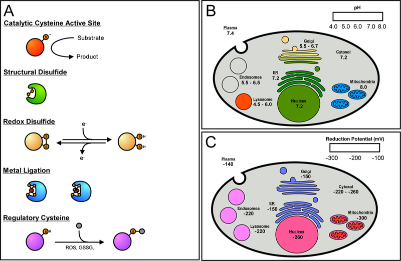
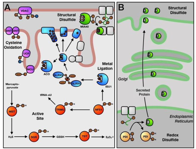
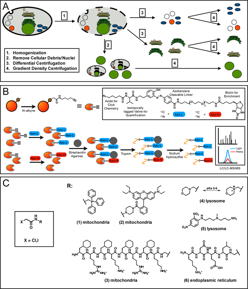
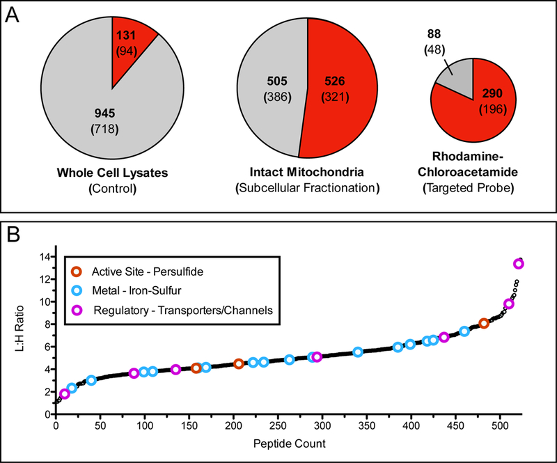
Cysteine residues are concentrated at key functional sites within proteins, performing diverse roles in metal binding, catalysis, and redox chemistry. Chemoproteomic platforms to interrogate the reactive cysteinome have developed significantly over the past 10 years, resulting in a greater understanding of cysteine functionality, modification, and druggability. Recently, chemoproteomic methods to examine reactive cysteine residues from specific subcellular organelles have provided significantly improved proteome coverage and highlights the unique functionalities of cysteine residues mediated by cellular localization. Here, the diverse physicochemical properties of the mammalian subcellular organelles are explored in the context of their effects on cysteine reactivity. The unique functions of cysteine residues found in the mitochondria and endoplasmic reticulum are highlighted, together with an overview into chemoproteomic platforms employed to investigate cysteine reactivity in subcellular organelles.


Figures

Figure 1:
Figure 1:
The functional roles of reactive cysteine residues and the pH and redox ranges of the mammalian organelle. (A) Cysteine residues can be grouped into several functional classes; catalytic, structural, redox-active, metal ligating, and regulatory. (B) The pH ranges found in the organelle of a typical mammalian cell. Red/orange is indicative of a low pH, green of a neutral pH, and blue of a higher, more alkaline pH. The mitochondrial pH is referring to the pH found in the mitochondrial matrix. (C) The reduction potentials found in the various organelles, where red indicates a low potential, and blue a higher relative reduction potential. The mitochondrial reduction potential refers to the mitochondrial matrix. The intermembrane space has a higher potential of ~ −255 mV.
Figure 2:
Figure 2:
Functional cysteine residues of the mitochondria and endoplasmic reticulum (ER). (A) Mitochondrial cysteine residues involved in persulfide chemistry (red -active site), iron-sulfur trafficking (blue - metal ligation), disulfide bond formation (green - structural disulfide), and oxidative regulation of membrane transporters (purple - regulatory). (B) ER cysteine residues involved in redox-dependent processes (yellow - redox disulfide).
Figure 3:
Figure 3:
Chemoproteomic platforms to study organelle-specific reactive and functional cysteine residues. (A) Enrichment and isolation of subcellular organelles is most commonly achieved through differential and gradient centrifugation. Cells are gently lysed by dounce homogenization (1), heavy cellular debris and intact nuclei are removed by low speed centrifugation (2), differential centrifugation at moderate and high-speed centrifugation can isolate fractions of organelle (3), which can be fully separated and enriched by density gradient centrifugation (4). (B) Cysteine residues from intact organelles can be quantified by labeling with IA-alkyne, followed by click-chemistry with a chemically cleavable biotin-azide tag. For quantification, an isotopic label is incorporated into either the protein (SILAC), the IA-alkyne probe, or the biotin-azide tag. Tagged proteins are enriched on streptavidin, reduced and alkylated, cleaved by trypsin on-bead, and labeled peptides released by chemical cleavage of the biotin-azide tag. These peptides are then analyzed by LC/LC-MS/MS, followed by isotopic quantification to determine peptide L/H ratios. (C) Cysteine-reactive groups can be appended to organelle-targeting groups. These groups include mitochondrial targeting lipophilic cations, such as rhodamine (1) and triphenylphosphonium (2), mitochondrial-targeting peptide, FxRFxKFxRFxK (3), lysosomal-targeting groups, such as morpholine (4) and 3-(2,4,-dinitroanilino)-3’-amino-N-methyldipropyl-amine (DAMP) (5), and ER-targeting peptide, KDEL (6)
Figure 4:
Figure 4:
Application of organelle-specific proteomics to examine mitochondrial reactive cysteine residues. (A) Pie charts demonstrate the fraction of mitochondrial peptides identified through probe labeling and proteomic analysis of whole cell lysates by iodoacetamide (IA)-alkyne (left), enriched mitochondria labeled with IA-alkyne (center) [65], and live cells labeled with rhodamine-chloroacetamide (right) [66]. Red shading indicates fraction of total peptides annotated as mitochondrial, while the gray shading indicates peptides from other subcellular organelle or those without annotation. Bold number indicates identified peptides, number within parentheses is the total number of proteins those peptides were identified from. (B) L/H ratios for mitochondrial cysteine-containing peptides identified by labeling of enriched mitochondria with either 100 μM (light) or 10 μM (heavy) IA-alkyne. Lower L/H ratios indicate more reactive cysteine residues. Colored open circles indicate cysteine residues that are annotated with functions described previously as important and somewhat specific to the mitochondria (red - active site cysteine persulfide, blue - iron-sulfur ligating, and pink - regulatory oxidation). Importantly these annotations are generally not observed during whole cell lysate analysis.

References

    1. Marino SM, Gladyshev VN: Cysteine function governs its conservation and degeneration and restricts its utilization on protein surfaces. J. Mol. Biol. 2010, 404:902–916. - PMC - PubMed
    1. Pe’er I, Felder CE, Man O, Silman I, Sussman JL, Beckmann JS: Proteomic signatures: amino acid and oligopeptide compositions differentiate among phyla. Proteins 2004, 54:20–40. - PubMed
    1. Pace NJ, Weerapana E: Diverse functional roles of reactive cysteines. ACS Chem. Biol. 2013, 8:283–296. - PubMed
    1. Weerapana E, Wang C, Simon GM, Richter F, Khare S, Dillon MBD, Bachovchin DA, Mowen K, Baker D, Cravatt BF: Quantitative reactivity profiling predicts functional cysteines in proteomes. Nature 2010, 468:790–795. - PMC - PubMed
    1. Bulaj G, Kortemme T, Goldenberg DP: Ionization-reactivity relationships for cysteine thiols in polypeptides. Biochemistry 1998, 37:8965–8972. - PubMed

Publication types

LinkOut - more resources