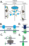
SOCE and STIM1 signaling in the heart: Timing and location matter
- PMID: 30508734
- PMCID: PMC9520449
- DOI: 10.1016/j.ceca.2018.11.008
SOCE and STIM1 signaling in the heart: Timing and location matter
Abstract
Store operated Ca2+ entry (SOCE) is an ancient and ubiquitous Ca2+ signaling pathway discovered decades ago, but the function of SOCE in human physiology is only now being revealed. The relevance of this pathway to striated muscle was solidified with the description of skeletal myopathies that result from mutations in STIM1 and Orai1, the two SOCE components. Here, we consider the evidence for STIM1 and SOCE in cardiac muscle and the sinoatrial node. We highlight recent studies revealing a role for STIM1 in cardiac growth in response to developmental and pathologic cues. We also review the role of STIM1 in the regulation of SOCE and Ca2+ store refilling in a non-Orai dependent manner. Finally, we discuss the importance of this pathway in ventricular cardiomyocytes where SOCE contribute to developmental growth and in pacemaker cells where SOCE likely has a fundamental to generating the cardiac rhythm.
Keywords: Cardiac muscle; Store-operated calcium channels; Store-operated calcium entry; Stromal interaction molecule 1 (STIM1).
Copyright © 2018 Elsevier Ltd. All rights reserved.
Figures


Similar articles
-
SOCE in the cardiomyocyte: the secret is in the chambers.Pflugers Arch. 2021 Mar;473(3):417-434. doi: 10.1007/s00424-021-02540-3. Epub 2021 Feb 27. Pflugers Arch. 2021. PMID: 33638008 Free PMC article. Review.
-
Filamin A Modulates Store-Operated Ca2+ Entry by Regulating STIM1 (Stromal Interaction Molecule 1)-Orai1 Association in Human Platelets.Arterioscler Thromb Vasc Biol. 2018 Feb;38(2):386-397. doi: 10.1161/ATVBAHA.117.310139. Epub 2017 Dec 28. Arterioscler Thromb Vasc Biol. 2018. PMID: 29284605
-
Interplay between ER Ca2+ Binding Proteins, STIM1 and STIM2, Is Required for Store-Operated Ca2+ Entry.Int J Mol Sci. 2018 May 19;19(5):1522. doi: 10.3390/ijms19051522. Int J Mol Sci. 2018. PMID: 29783744 Free PMC article.
-
Role of STIM1/ORAI1-mediated store-operated Ca2+ entry in skeletal muscle physiology and disease.Cell Calcium. 2018 Dec;76:101-115. doi: 10.1016/j.ceca.2018.10.004. Epub 2018 Oct 30. Cell Calcium. 2018. PMID: 30414508 Free PMC article. Review.
-
STIM-TRP Pathways and Microdomain Organization: Contribution of TRPC1 in Store-Operated Ca2+ Entry: Impact on Ca2+ Signaling and Cell Function.Adv Exp Med Biol. 2017;993:159-188. doi: 10.1007/978-3-319-57732-6_9. Adv Exp Med Biol. 2017. PMID: 28900914 Review.
Cited by
-
Potential Roles of IP3 Receptors and Calcium in Programmed Cell Death and Implications in Cardiovascular Diseases.Biomolecules. 2024 Oct 20;14(10):1334. doi: 10.3390/biom14101334. Biomolecules. 2024. PMID: 39456267 Free PMC article. Review.
-
Ca2+ Signaling in Cardiac Fibroblasts and Fibrosis-Associated Heart Diseases.J Cardiovasc Dev Dis. 2019 Sep 23;6(4):34. doi: 10.3390/jcdd6040034. J Cardiovasc Dev Dis. 2019. PMID: 31547577 Free PMC article. Review.
-
Cardiomyocyte-Specific Deletion of Orai1 Reveals Its Protective Role in Angiotensin-II-Induced Pathological Cardiac Remodeling.Cells. 2020 Apr 28;9(5):1092. doi: 10.3390/cells9051092. Cells. 2020. PMID: 32354146 Free PMC article.
-
TRIC-A regulates intracellular Ca2+ homeostasis in cardiomyocytes.Pflugers Arch. 2021 Mar;473(3):547-556. doi: 10.1007/s00424-021-02513-6. Epub 2021 Jan 21. Pflugers Arch. 2021. PMID: 33474637 Free PMC article. Review.
-
Side-by-side comparison of published small molecule inhibitors against thapsigargin-induced store-operated Ca2+ entry in HEK293 cells.PLoS One. 2024 Jan 23;19(1):e0296065. doi: 10.1371/journal.pone.0296065. eCollection 2024. PLoS One. 2024. PMID: 38261554 Free PMC article.
References
-
- Yaniv Y, Sirenko S, Ziman BD, Spurgeon HA, Maltsev VA, Lakatta EG, New evidence for coupled clock regulation of the normal automaticity of sinoatrial nodal pacemaker cells: bradycardic effects of ivabradine are linked to suppression of intracellular Ca(2)(+) cycling, Journal of molecular and cellular cardiology, 62 (2013) 80–89. - PMC - PubMed
-
- Heineman FW, Balaban RS, Control of mitochondrial respiration in the heart in vivo, Annu Rev Physiol, 52 (1990) 523–542. - PubMed
Publication types
MeSH terms
Substances
Grants and funding
LinkOut - more resources
Full Text Sources
Miscellaneous

