Review
. 2018:75:1-213.
doi: 10.1016/bs.accb.2018.09.001. Epub 2018 Nov 28.

Exploration of the Sialic Acid World

Affiliations
Review

Exploration of the Sialic Acid World

Roland Schauer et al. Adv Carbohydr Chem Biochem. 2018.

Abstract

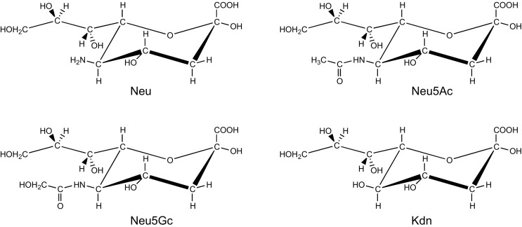
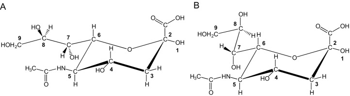
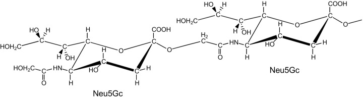
Sialic acids are cytoprotectors, mainly localized on the surface of cell membranes with multiple and outstanding cell biological functions. The history of their structural analysis, occurrence, and functions is fascinating and described in this review. Reports from different researchers on apparently similar substances from a variety of biological materials led to the identification of a 9-carbon monosaccharide, which in 1957 was designated "sialic acid." The most frequently occurring member of the sialic acid family is N-acetylneuraminic acid, followed by N-glycolylneuraminic acid and O-acetylated derivatives, and up to now over about 80 neuraminic acid derivatives have been described. They appeared first in the animal kingdom, ranging from echinoderms up to higher animals, in many microorganisms, and are also expressed in insects, but are absent in higher plants. Sialic acids are masks and ligands and play as such dual roles in biology. Their involvement in immunology and tumor biology, as well as in hereditary diseases, cannot be underestimated. N-Glycolylneuraminic acid is very special, as this sugar cannot be expressed by humans, but is a xenoantigen with pathogenetic potential. Sialidases (neuraminidases), which liberate sialic acids from cellular compounds, had been known from very early on from studies with influenza viruses. Sialyltransferases, which are responsible for the sialylation of glycans and elongation of polysialic acids, are studied because of their significance in development and, for instance, in cancer. As more information about the functions in health and disease is acquired, the use of sialic acids in the treatment of diseases is also envisaged.

Keywords: History; Sialic acids; Sialobiochemistry; Sialobiology; Sialochemistry.


Figures

Fig. 1
Fig. 1
The “mother” molecule of the family Sialic Acids (Neu), together with the three major “children” (Neu5Ac, Neu5Gc, Kdn), presented in the α-configuration, as occurring in sialic acid-containing carbohydrate chains.
Fig. 2
Fig. 2
Chemical structures of N-acetylneuraminic acid (5-acetamido-3,5-dideoxy-d-glycero-d-galacto-non-2-ulosonic acid, Neu5Ac) in different views according to IUPAC/IUBMB recommendations. (A) Neu5Ac in Fischer projection formula, open chain; (B) β-Neu5Ac/N-acetyl-β-neuraminic acid/5-acetamido-3,5-dideoxy-d-glycero-β-d-galacto-non-2-ulopyranosonic acid in Fischer projection formula, cyclic chain, pyranose ring; (C) β-Neu5Ac in Haworth representation, pyranose ring; and (D) β-Neu5Ac in 2C5 chair conformation.
Fig. 3
Fig. 3
Chemical structures of (A) Neu1,5lactam (5,2B conformation), (B) Neu2en5Ac (4H5 conformation), (C) CMP-β-Neu5Ac, (D) Neu2,7an5Ac (5C2 conformation), (E) Neu5Ac1,7lactone (5C2 conformation), and (F) Neu4,8an5Ac (7C4 conformation; two tautomers).
Fig. 4
Fig. 4
(A) Gunnar Blix (1894–1981), (B) Ernst Klenk (1896–1971), (C) Alfred Gottschalk (1894–1973).
Fig. 5
Fig. 5
IR spectra of “N-acetylneuraminic acid,” enzymatically released (V. cholerae) from (1) human urinary mucoprotein, (2) bovine submaxillary gland mucin (crystallization fraction I), and (3) bovine submaxillary gland mucin (crystallization fraction II), and chemically released from (4) bovine submaxillary gland mucin.
Fig. 6
Fig. 6
X-ray powder diagrams of “sialic acid” isolated from submaxillary gland mucins [(A) bovine, (B) ovine, (C) porcine, (D) equine] and “methoxyneuraminic acid” (E). Guinier camera. Nickel-filtered copper K-radiation.
Scheme 1
Scheme 1
Proposed structure of “Kohlenhydrat I” with the formation of pyrrole-2-carboxylic acid (and d-erythrose), according to Hiyama, taking into account Blix's findings.,
Fig. 7
Fig. 7
Structure proposal of Yamakawa and Suzuki for “hemataminic acid” (C10H19NO8), a methoxy derivative of a 2-amino-2-deoxy-nonuronic acid.
Fig. 8
Fig. 8
Gottschalk's N-glycosyl-pyrrole-2-carboxylic acid proposal (A, free form; B, bound form) for the structure of the enzymatically released substance from mucoprotein.,
Scheme 2
Scheme 2
Gottschalk's O-glycosyl-pyrrole-2-carboxylic acid proposals for the structure of the (bio)chemically released substance from mucoprotein with the formation of pyrrole-2-carboxylic acid.,
Scheme 3
Scheme 3
Reaction scheme to explain Gottschalk's proposal for the structures of “sialic acid” (a and b) (C13H21NO10), “neuraminic acid” (c), and “methoxyneuraminic acid” (f, C10H19NO8), including the formation of pyrrole-2-carboxylic acid (d) and the tetrose (e), and a possible intermediate g.,
Fig. 9
Fig. 9
Structure of “methoxyneuraminic acid” according to Klenk and coworkers; C11H21NO9 (mw 311) was the best fit with the elemental analysis. Note that the ultimately correct chemical formula, C10H19NO8 (mw 281), was pushed aside. Interestingly, molecular weight determination, via cryoscopy in water, yielded 273 and 276 Da.
Fig. 10
Fig. 10
Survey of the search for the stereochemistry around C4, C5, C6, C7, and C8 of N-acetylneuraminic acid in a historical perspective: a path of trial and error (see Section 4).
Scheme 4
Scheme 4
Synthesis of N-acetylneuraminic acid (Structure I; Fig. 10) from N-acetyl-d-glucosamine and oxaloacetic acid.
Fig. 11
Fig. 11
Saul Roseman (1921–2011).
Scheme 5
Scheme 5
N-Acetylneuraminic acid aldolase-catalyzed cleavage and synthesis of N-acetylneuraminic acid (Structure II; Fig. 10) with N-acetyl-d-mannosamine and pyruvic acid as counter compounds.,
Scheme 6
Scheme 6
Synthesis of N-acetylneuraminic acid (Structure II; Fig. 10) from d-GlcNAc or d-ManNAc and oxaloacetic acid.,
Scheme 7
Scheme 7
Chemical reactions to search for the chirality of the C4 atom of N-acetylneuraminic acid: The OH group at C4 should project to the left in the Fischer projection formula (Structure III; Fig. 10).
Scheme 8
Scheme 8
Three routes to prove the correct chirality around C4 of N-acetylneuraminic acid: The OH group at C4 should project to the right in the Fischer projection formula (Structure IV; Fig. 10).
Scheme 9
Scheme 9
Synthesis of 3-acetamido-3-deoxy-d-glycero-d-galacto-heptose from the γ-lactone of N-acetylneuraminic acid (Structure IV; Fig. 10).
Fig. 12
Fig. 12
Conclusive evidence for the 2C5 chair conformation of the pyranose ring, and the anomeric configuration assignment, of N-acetylneuraminic acid methyl ester (derivatives), based on 100-MHz 1H NMR analysis.
Scheme 10
Scheme 10
Elucidation of the stereochemistry around the anomeric carbon atom via a search for lactone formation, by subjecting the two anomeric N-acetylneuraminic acid methyl ester methyl glycosides to sequential periodate degradation, borohydride reduction, and treatment with N,N′-dicyclohexylcarbodiimide (DCC). The two starting compounds differ, after de-esterification, in their susceptibility to sialidase.
Fig. 13
Fig. 13
(A) Polarographic measurement of the periodic acid consumption at 0°C of isomeric N-acetyl-mono-O-acetyl-neuraminic acids. Neu5Ac, cross; Neu4,5Ac2, open circle; Neu5,7Ac2, closed circle; Neu5,8Ac2, closed square. (B) Expected cleavages in the C7–C8–C9 glycerol side chains are indicated with ------.
Fig. 14
Fig. 14
(A) EI mass spectrometric cleavages in the C7–C8–C9 glycerol side chain of N,O-acylneuraminic acids. (B) Fragmentation scheme for the analysis of sialic acid derivatives., Ac, acetyl; Lt, lactyl; Me, methyl; TMS, trimethylsilyl.
Fig. 15
Fig. 15
(A) EI mass spectrum (70 eV) of the per-O-trimethylsilylated methyl ester of β-Neu5Ac. (B) EI mass spectrum (70 eV) of the per-O-trimethylsilylated methyl ester of β-Neu5,9Ac2.
Fig. 16
Fig. 16
Molecular conformation of crystalline α-Neu5Ac methyl ester methyl glycoside.
Fig. 17
Fig. 17
60-MHz 1H NMR spectrum of Neu5Ac, dissolved in D2O, recorded at ambient temperature.
Fig. 18
Fig. 18
500-MHz 1H NMR spectrum of Neu5Ac (β:α = 93:7), dissolved in D2O, recorded at pD 7 and 27°C (Table 3), relative to internal acetone in D2O (δ 2.225 ppm). The H3e and H3a signals of α-Neu5Ac are enlarged 10-fold.
Fig. 19
Fig. 19
Proton-noise-decoupled 25.2-MHz 13C NMR spectrum of Neu5Ac, dissolved in D2O, recorded at pD ~2 and 25°C, relative to external TMS (δ 0 ppm). Only the signals stemming from the β-anomer are clearly observable. For a first report on chemical shifts of both α- and β-Neu5Ac, see ref. .
Fig. 20
Fig. 20
NMR Molecular conformations in aqueous solution of (A) β-Neu5Ac according to ref. , (B) β-Neu5Ac according to refs. , , and (C) and (D) α-Neu5Ac according to refs. , . For both (A) and (B), as measured in D2O solution, the H atoms of the OH and NH groups would be replaced by D atoms.
Fig. 21
Fig. 21
Ab initio calculated structure of α-Neu5Ac.
Fig. 22
Fig. 22
(A, B) Two different drawings of the structure of α-Neu5Ac.
Scheme 11
Scheme 11
Possible pyrrole chromogen formation [N-deacetylation, condensation NH2(C5) and CO(C2), decarboxylation, dehydration] in the Bial (orcinol/concd. HCl/FeCl3) and Svennerholm (resorcinol/concd. HCl/CuSO4) reaction, taking into account the proposals in refs. and .
Scheme 12
Scheme 12
Possible pyrrole derivatives arising from Neu5Ac, following the “direct” Ehrlich (E = p-dimethylaminobenzaldehyde/concd. HCl) and the “indirect” Ehrlich (alkali; E = p-dimethylaminobenzaldehyde/concd. HCl) reaction, taking into account the proposals in refs. and , , .
Scheme 13
Scheme 13
Proposed reaction mechanisms of the periodic acid/thiobarbituric acid assay., , ,
Scheme 14
Scheme 14
Proposed reaction mechanism for the transamination of sialic acid with pyridoxamine in the presence of zinc acetate and pyridine/methanol, yielding the fluorescent Zn-chelate.
Scheme 15
Scheme 15
Reaction of formaldehyde with acetylacetone in the presence of ammonium acetate, yielding the fluorigen 3,5-diacetyl-1,4-dihydro-2,6-dimethylpyridine.,
Fig. 23
Fig. 23
TLC patterns of sialic acids on a cellulose plate, developed with propan-1-ol/butan-1-ol/0.1 M HCl (2:1:1, v/v/v), and stained with Bial's reagent. Lane 1: sialic acid mixture, released from bovine submandibular gland mucin. Lane 2: standard Neu5Ac and Neu5Gc.
Scheme 16
Scheme 16
Reaction of Neu5Ac with 1,2-diamino-4,5-methylenedioxybenzene (DMB) to yield the corresponding fluorescent derivative.,
Fig. 24
Fig. 24
Detection of sialic acid residues in human colon by silver-intensified lectin–gold labeling of sections from paraffin-embedded tissues. (A) Normal colonic surface (arrowhead) and crypt epithelium (asterisks) are reactive for (α2→3)-linked sialic acid residues, as detected with the Maackia amurensis lectin. (B) In contrast, (α2→6)-linked sialic acid residues are undetectable in the colonic surface (arrowhead) and crypt epithelium (asterisks) with the Sambucus nigra lectin. Note the positive labeling of capillary endothelia (arrows). In two adjacent serial sections of a colon carcinoma, both (α2→3)-linked sialic acid residues (C) and (α2→6)-linked sialic acid residues (D) can be detected. Scale bar: 20 μm (A, B), 50 μm (C, D).
Fig. 25
Fig. 25
Reexpression of (α2→8)-linked polysialic acid in human Wilms tumor. (A) Silver-intensified immunogold staining with mAb 735 of a section of paraffin-embedded tissue shows intense labeling in the Wilms tumor (WT). The adjacent normal kidney (NK) is unlabeled. (B) Immunolabeling in a consecutive serial section for (α2→8)-linked polysialic acid is abolished by section pretreatment with endoneuraminidase N (Endo N). (C) Immunogold labeling for (α2→8)-linked polysialic acid of an ultrathin frozen section from a Wilms tumor is very intense at cell-surface regions with a thick surface coat. (D) In contrast, immunogold labeling is sparse and patchy in regions of close contact of tumor cells. Scale bar: 300 μm (A, B), 300 nm (C), 150 nm (D).
Fig. 26
Fig. 26
A terminal Neu5Gc(α2→O5)Neu5Gc sequence.
Fig. 27
Fig. 27
2C5 Chair conformations of α-Pse, β-Leg, β-4eLeg, β-8eLeg, α-Aci, and α-8eAci. Note the difference in orientation of the COOH and OH groups at the anomeric C2 atom.
Fig. 28
Fig. 28
Metabolism of non-2-ulosonic acids having the d-glycero-d-galacto configuration. (A) N-Acetylneuraminic acid (Neu5Ac) in vertebrates; (B) N-acetylneuraminic acid (Neu5Ac) in bacteria; (C) ketodeoxynononic acid (Kdn) in vertebrates; and (D) 5,7-di-N-acetyllegionaminic acid (Leg5,7Ac2) in bacteria.d-6dManNAc4NAc, 2,4-diacetamido-2,4,6-trideoxy-d-mannose; GDP-α-d-6dGlcNAc4NAc, GDP-2,4-diacetamido-2,4,6-trideoxy-α-d-glucose; Kdn9P, ketodeoxynononic acid 9-phosphate; d-ManNAc6P, N-acetyl-d-mannosamine 6-phosphate; d-Man6P, d-mannose 6-phosphate; Neu5Ac9P, N-acetylneuraminic acid 9-phosphate; PEP, phosphoenolpyruvate; Pi, phosphate; PPi, diphosphate. In case of CMP-β-Leg5,7Ac2, also another route, going from UDP-α-d-GlcNAc, via UDP-α-d-6dGlcNAc4NAc to d-6dManNAc4NAc, has been formulated.
Fig. 29
Fig. 29
Time plots for the migration of O-acetyl groups in (A) Neu5,7Ac2 (α:β = 23:77) → Neu5,9Ac2 (α:β = 9:91) and (B) Neu5,7,9Ac3 (α:β = 22:78) → Neu5,8,9Ac3 (α:β = 3:97) in 0.1 M phosphate, pD 7.2–7.5, at 37°C, as monitored by 360-MHz 1H NMR spectroscopy. As a result of the migration, an anomerization process will also occur.
Fig. 30
Fig. 30
Metabolism of sialic acid O-acetylation (here Neu5Ac), including a survey of enzymes involved in the transfer and removal of O-acetyl groups. The flowers indicate the hydroxy groups found to be O-acetylated. The hands symbolize the position specificity of the O-acetyltransferase activities discovered: sialate-4-O-acetyltransferase, sialate-7-O-acetyltransferase, and sialate-9-O-acetyltransferase. The scissors represent the sialate-4-O-acetylesterase and sialylate-9-O-acetylesterase, involved in the hydrolysis of the ester groups. The O-acetyl group at the sialic acid glycerol side chain can migrate between C7 and C9 (via C8?), whereas the 4-O-acetyl group seems to be immobile.
Fig. 31
Fig. 31
Survey of naturally occurring substituents of the sialic acid backbone. For a full survey of structures, see Table 1.
Fig. 32
Fig. 32
(A) exo- and endo-Sialidase activities. (B) trans-Sialidase activities.
Fig. 33
Fig. 33
Structures of (A) Relenza® and (B) Tamiflu®.
Fig. 34
Fig. 34
Binding of sialidase-treated rat erythrocytes, lymphocytes, and thrombocytes to homologues rat peritoneal macrophages by the galactose-specific acceptor. (A) Scanning electron microscopy of erythrocyte–macrophage interaction. After prolonged incubation, the erythrocytes were ingested; (B) scanning electron microscopy of cultured, sialidase-treated lymphocytes bound as a rosette by a peritoneal macrophage; (C) scanning electron microscopy of the characteristic binding of a single sialidase-treated lymphocyte by a rat peritoneal macrophage. In contrast to sialidase-treated, bound erythrocytes, no deformation of phagocytosis is observed. (D) Scanning electron microscopy of sialidase-treated thrombocytes to a macrophage adherent to a Petri dish.
Fig. 35
Fig. 35
Lectins, for example, the sialic acid-binding Sambucus nigra agglutinin (SNA), protect plants against herbivorous and chewing animals.
Fig. 36
Fig. 36
The selectins play a key role in the control of leukocyte traffic in the body. (A) L-Selectin functions in the migration of lymphocytes to lymphoid organs. (B) E- and P-Selectins mediate the recruitment of neutrophils to sites of inflammation.

References

    1. Montreuil J., Vliegenthart J.F.G., Schachter H. In: Glycoproteins II. Montreuil J., Vliegenthart J.F.G., Schachter H., editors. Vol. 29b. Elsevier Science B.V; Amsterdam, The Netherlands: 1997. Preface; pp. v–vi. (New Comprehensive Biochemistry).
    1. Warren L. The Distribution of Sialic Acids in Nature. Comp. Biochem. Physiol. 1963;10:153–171. - PubMed
    1. Warren L. In: Biological Roles of Sialic Acid. Rosenberg A., Schengrund C.-L., editors. Plenum Press; New York, NY, USA: 1976. The Distribution of Sialic Acids Within the Eukaryotic Cell; pp. 103–121.
    1. Ng S.-S., Dain J.A. In: Biological Roles of Sialic Acid. Rosenberg A., Schengrund C.-L., editors. Plenum Press; New York, NY, USA: 1976. The Natural Occurrence of Sialic Acids; pp. 59–102.
    1. Corfield A.P., Schauer R. In: Sialic Acids—Chemistry, Metabolism and Function. Schauer R., editor. Vol. 10. Springer-Verlag; Vienna, Austria: 1982. Occurrence of Sialic Acids; pp. 5–50. (Cell Biology Monographs).

MeSH terms

Substances

Supplementary concepts