. 2019 Jul:25:101051.
doi: 10.1016/j.redox.2018.11.009. Epub 2018 Nov 17.

GPx3 supports ovarian cancer progression by manipulating the extracellular redox environment

Affiliations

GPx3 supports ovarian cancer progression by manipulating the extracellular redox environment

Beth L Worley et al. Redox Biol. 2019 Jul.

Abstract

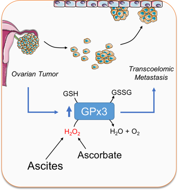
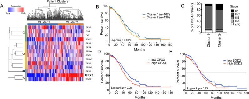
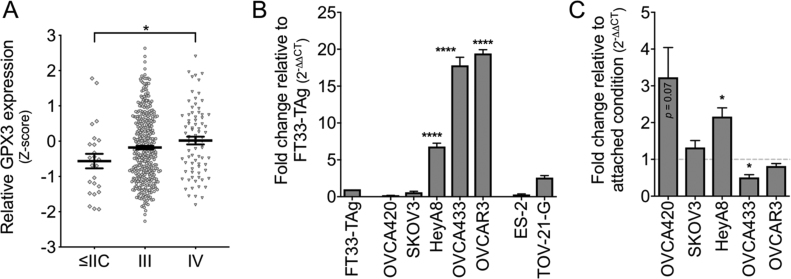
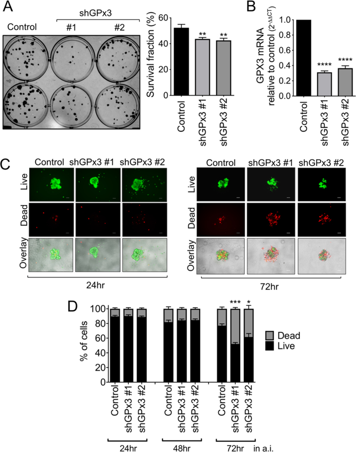
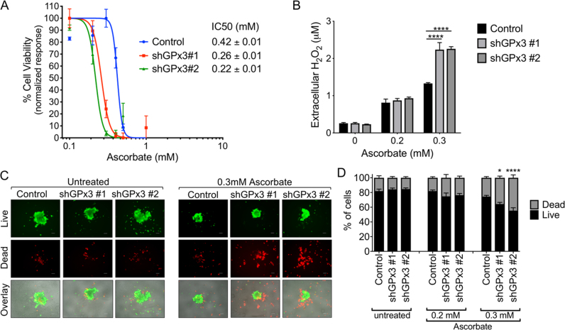
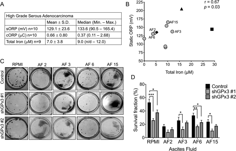
Ovarian cancer remains the most lethal gynecologic malignancy, and is primarily diagnosed at late stage when considerable metastasis has occurred in the peritoneal cavity. At late stage abdominal cavity ascites accumulation provides a tumor-supporting medium in which cancer cells gain access to growth factors and cytokines that promote survival and metastasis. However, little is known about the redox status of ascites, or whether antioxidant enzymes are required to support ovarian cancer survival during transcoelomic metastasis in this medium. Gene expression cluster analysis of antioxidant enzymes identified two distinct populations of high-grade serous adenocarcinomas (HGSA), the most common ovarian cancer subtype, which specifically separated into clusters based on glutathione peroxidase 3 (GPx3) expression. High GPx3 expression was associated with poorer overall patient survival and increased tumor stage. GPx3 is an extracellular glutathione peroxidase with reported dichotomous roles in cancer. To further examine a potential pro-tumorigenic role of GPx3 in HGSA, stable OVCAR3 GPx3 knock-down cell lines were generated using lentiviral shRNA constructs. Decreased GPx3 expression inhibited clonogenicity and anchorage-independent cell survival. Moreover, GPx3 was necessary for protecting cells from exogenous oxidant insult, as demonstrated by treatment with high dose ascorbate. This cytoprotective effect was shown to be due to GPx3-dependent removal of extracellular H2O2. Importantly, GPx3 was necessary for clonogenic survival when cells were cultured in patient-derived ascites fluid. While oxidation reduction potential (ORP) of malignant ascites was heterogeneous in our patient cohort, and correlated positively with ascites iron content, GPx3 was required for optimal survival regardless of ORP or iron content. Collectively, our data suggest that HGSA ovarian cancers cluster into distinct groups of high and low GPx3 expression. GPx3 is necessary for HGSA ovarian cancer cellular survival in the ascites tumor environment and protects against extracellular sources of oxidative stress, implicating GPx3 as an important adaptation for transcoelomic metastasis.

Keywords: Ascites; Ascorbate; Extracellular glutathione peroxidase; GPx3; Ovarian cancer.


Figures

fx1
Graphical abstract
Fig. 1
Fig. 1
Ovarian cancer specimens fall into two distinct clusters based on expression of the extracellular antioxidant GPx3. A: Hierarchical cluster analysis of antioxidant gene expression separates high grade serous adenocarcinoma patient samples into two distinct clusters (TCGA RNA sequencing data log2 transformed). B: Kaplan-Meier analysis of overall survival cluster 1 and cluster 2 patients (Log-rank Mantel-Cox test, HR: 1.20 [95% CI: 0.90–1.61]). C: Percentages of different tumor stages at diagnosis of patients in cluster 1 and cluster 2. D: High GPx3 expression is negatively associated with patient survival. TCGA RNA sequencing samples were sorted above (high) and below (low) median GPx3 expression, and stratified in Kaplan Meier plot by overall survival (Log-rank Mantel-Cox test, HR: 1.30 [95% CI: 0.97–1.75]). E: Kaplan Meier plot comparing overall survival of patients with high and low SOD2 expression. (Log-rank Mantel-Cox test, HR: 1.03 [95% CI: (0.89–1.60]).
Fig. 2
Fig. 2
GPx3 expression correlates with tumor stage and is increased in several HGSA ovarian cancer cell lines. A: GPx3 mRNA expression (TCGA, z-scores) in HGSA specimens stratified by tumor stage (n = 24, stage p = 0.03, Tukey's post hoc * p < 0.05). B: GPx3 mRNA expression relative to normal FT33-TAg cells was assessed by semi-quantitative real time RT-PCR mean ± SEM (n = 3; ANOVA p < 0.0001, Tukey's post hoc **** p < 0.0001). C: GPx3 expression in response to anchorage independent culturing in ULA plates (24 h), expressed relative to GPx3 expression in same cell line under attached conditions (mean ± SEM, n = 3; t-test relative to attached conditions * p < 0.05).
Fig. 3
Fig. 3
GPx3 knockdown inhibits OVCAR3 clonogenicity and survival in anchorage independence. A: Cells (100/well) were seeded onto 12-well plates and stained with crystal violet after 10 days of growth. Data expressed as mean ± SEM of 3 independent experiments (n = 4/group; ANOVA, post hoc **p< 0.01). B: GPx3 knockdown by shRNA was verified by semi quantitative real time RT-PCR. C: The effects of GPx3 knockdown on cell viability were assessed under conditions of anchorage independence (a.i.), by culturing cells in 96 well ultra-low attachment (ULA; 1000 cells/well) plates for indicated times, followed by staining with calcein-AM (2 μM), indicating live cells, and Ethidium homodimer-1 (4 μM) indicating dead cells. Representative images are shown. Scale bar = 100 µm. D: Quantification of live/dead staining using ImageJ. Data expressed as mean ± SEM (n = 4–8/group; One-way ANOVA, Tukey's post hoc, **p< 0.01, ****p < 0.0001).
Fig. 4
Fig. 4
GPx3 knockdown increases cell death and levels of extracellular H2O2 in response to high dose ascorbate. A: IC50 values of ascorbate on ovarian cancer cells were determined by cell viability assay. OVCAR3 shGPx3 stable cell lines (5 × 103/well) were seeded into 96-well plates and exposed to various concentrations of ascorbate for 24 h. Cell viability was determined by crystal violet uptake and IC50 values calculated as shown. B: GPx3 scavenges ascorbate-induced H2O2. Cells (5 ×103/well) were seeded into 96-well plates and treated with ascorbate for 24 h. Medium from each well was collected and immediately analyzed using Amplex Red·H2O2 concentrations were derived from standard curves and data expressed as mean ± SEM (n = 3/group; Two-way ANOVA, Tukey's post hoc, ****p< 0.0001). C: The effects of GPx3 knockdown on cell viability were assessed under conditions of anchorage independence by culturing cells in ULA plates and treated with 0.3 mM ascorbate for 48 h, followed by staining with calcein-AM to indicate live cells (green), and Ethidium homodimer-1 to indicate dead cells (red; Bottom image overlay). Representative images are shown. Scale bar = 100 µm. D: Quantification of live/dead cell fractions in anchorage-independent OVCAR3 spheroids following 48 h treatment with indicated doses of ascorbate. Data expressed as mean ± SEM form 3 independent experiments (n = 4–12/group; one-way ANOVA, Tukey's post hoc, * p < 0.05, ****p< 0.0001).
Fig. 5
Fig. 5
GPx3 is required for cell survival in ascites. A: Summary of static (sORP), capacity oxidation reduction potential (cORP) and iron content of HGSA derived ascites. Ascites were cleared of all cells by centrifugation and filtration and sORP and capacity cORP were measured using the RedoxSYS. Total iron was measured using a colorimetric assay. B: Positive correlation between ascites sORP and iron content. Ascites were derived from high grade serous ovarian adenocarcinomas (〇) granulosa tumor (♦), endometrial ovarian cancer (▲), GI tumor (■; non-parametric Spearman correlation). C: Clonogenicity of OVCAR3 control or GPx3 shRNA knock-down cells (45 cells/well in 24 well plate) were seeded in ascites fluids with high (AF 15) and median sORP (AF 2, 3, 6). Clonogenicity was quantified after 10 days as in Fig. 3. Images are representative of at least 3 independent assays. Data expressed as mean ± SEM (n = 4/group, ANOVA, Tukey's post hoc **p < 0.05, **p < 0.01, ***p < 0.001). D: Quantification of clonogenicity using ImageJ.

References

    1. Torre L.A., Trabert B., DeSantis C.E., Miller K.D., Samimi G., Runowicz C.D., Gaudet M.M., Jemal A., Siegel R.L. Ovarian cancer statistics, 2018. CA Cancer J. Clin. 2018;68(4):284–296. - PMC - PubMed
    1. Davidson B. Ovarian carcinoma and serous effusions. Changing views regarding tumor progression and review of current literature. Anal. Cell.Pathol. J. Eur. Soc. Anal. Cell. Pathol. 2001;23(3–4):107–128. - PMC - PubMed
    1. Latifi A., Luwor R.B., Bilandzic M., Nazaretian S., Stenvers K., Pyman J., Zhu H., Thompson E.W., Quinn M.A., Findlay J.K., Ahmed N. Isolation and characterization of tumor cells from the ascites of ovarian cancer patients: molecular phenotype of chemoresistant ovarian tumors. PLoS One. 2012;7(10):e46858. - PMC - PubMed
    1. Tan D.S., Agarwal R., Kaye S.B. Mechanisms of transcoelomic metastasis in ovarian cancer. Lancet Oncol. 2006;7(11):925–934. - PubMed
    1. Piskounova E., Agathocleous M., Murphy M.M., Hu Z., Huddlestun S.E., Zhao Z., Leitch A.M., Johnson T.M., DeBerardinis R.J., Morrison S.J. Oxidative stress inhibits distant metastasis by human melanoma cells. Nature. 2015;527(7577):186–191. - PMC - PubMed

Publication types