. 2019 Jan 29;63(2):e01439-18.
doi: 10.1128/AAC.01439-18. Print 2019 Feb.

Antibiotics Stimulate Formation of Vesicles in Staphylococcus aureus in both Phage-Dependent and -Independent Fashions and via Different Routes

Affiliations

Antibiotics Stimulate Formation of Vesicles in Staphylococcus aureus in both Phage-Dependent and -Independent Fashions and via Different Routes

Federica Andreoni et al. Antimicrob Agents Chemother. .

Abstract

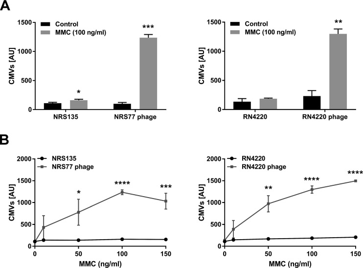
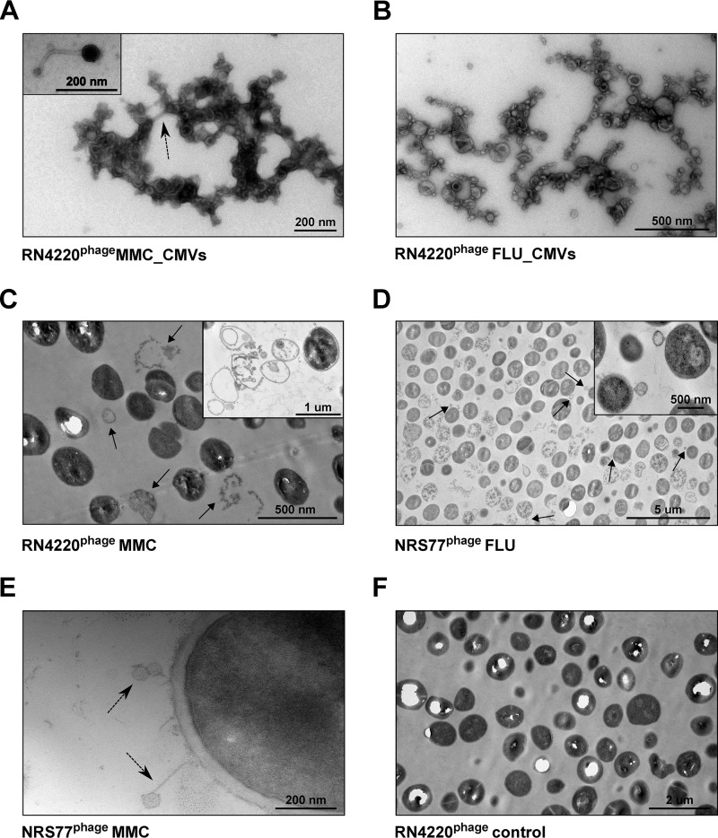
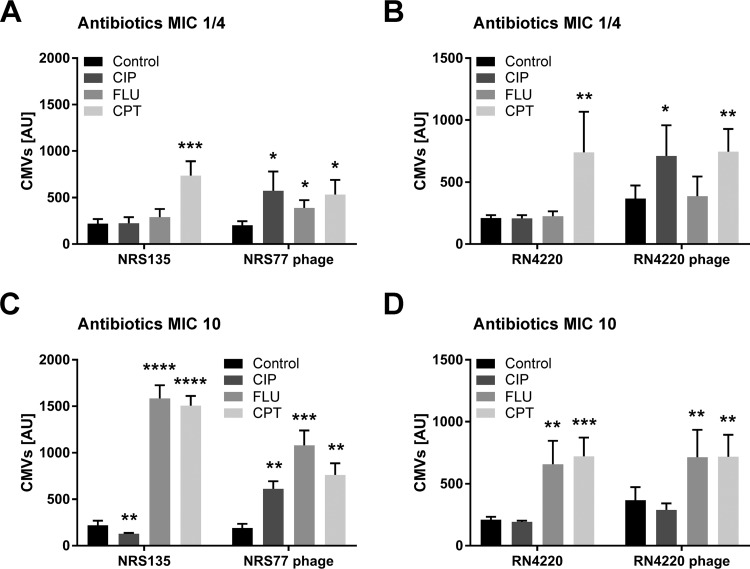
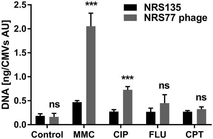
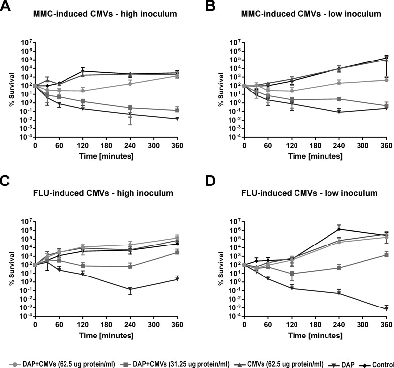
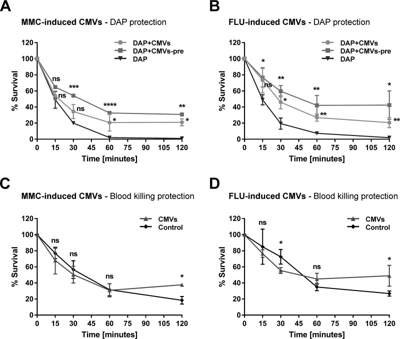
Bacterial membrane vesicle research has so far focused mainly on Gram-negative bacteria. Only recently have Gram-positive bacteria been demonstrated to produce and release extracellular membrane vesicles (MVs) that contribute to bacterial virulence. Although treatment of bacteria with antibiotics is a well-established trigger of bacterial MV formation, the underlying mechanisms are poorly understood. In this study, we show that antibiotics can induce MVs through different routes in the important human pathogen Staphylococcus aureus DNA-damaging agents and antibiotics inducing the SOS response triggered vesicle formation in lysogenic strains of S. aureus but not in their phage-devoid counterparts. The β-lactam antibiotics flucloxacillin and ceftaroline increased vesicle formation in a prophage-independent manner by weakening the peptidoglycan layer. We present evidence that the amount of DNA associated with MVs formed by phage lysis is greater than that for MVs formed by β-lactam antibiotic-induced blebbing. The purified MVs derived from S. aureus protected the bacteria from challenge with daptomycin, a membrane-targeting antibiotic, both in vitro and ex vivo in whole blood. In addition, the MVs protected S. aureus from killing in whole blood, indicating that antibiotic-induced MVs function as a decoy and thereby contribute to the survival of the bacterium.

Keywords: Staphylococcus aureus; antibiotics; bacteriophages; membrane vesicles.


Figures

FIG 1
FIG 1
MMC treatment induces CMV formation in lysogenic S. aureus strains NRS and RN4220. Mitomycin C (MMC) was added at 100 ng/ml (A) or at the concentrations indicated (B) to log-phase cultures of bacterial strains NRS135 and RN4220, as well as their lysogenic variants NRS77phage and RN4220phage. After 4 h of incubation, the amounts of CMVs in the supernatants were measured using a fluorescent dye and were quantified in arbitrary units (AU). In our system, a value of 1,500 AU corresponds to a CMV preparation containing 250 μg protein/ml. The graphs show data from at least three independent experiments. Statistical analysis was carried out using the unpaired t test. *, P < 0.05; **, P < 0.01; ***, P < 0.001; ****, P < 0.0001.
FIG 2
FIG 2
TEM images of membrane vesicles and S. aureus after stimulation with MMC or FLU. (A through E) TEMs of CMVs and cells of S. aureus RN4220phage and NRS77phage treated with either 100 ng/ml MMC (A, C, E) or 10 times the MIC of FLU (B and D). Phages are indicated by dashed arrows. In samples of MMC-treated cultures, the presence of ghost cells (C) (indicated by arrows and shown in the inset) and of phages (E) was observed. In cultures treated with FLU, high numbers of ghost cells and cells with blebs (indicated by arrows and shown in the inset) were observed (D). (F) Untreated RN4220phage cells.
FIG 3
FIG 3
Antibiotics triggered phage-dependent and -independent CMV induction in S. aureus strains NRS135 and RN4220. Log-phase cultures of strains NRS135 and RN4220, as well as their lysogenic variants NRS77phage and RN4220phage, were treated with ciprofloxacin (CIP), flucloxacillin (FLU), or ceftaroline (CPT) at concentrations equivalent to ¼ of the MIC (A and B) or 10× MIC (C and D). The amounts of CMVs in the supernatants were measured after 6 h of induction using a fluorescent dye and were quantified in arbitrary units (AU). In our system, a value of 1,500 AU corresponds to a CMV preparation containing 250 μg protein/ml. The graphs show data from at least three independent experiments. Statistical analysis was carried out using the unpaired t test. *, P < 0.05; **, P < 0.01; ***, P < 0.001; ****, P < 0.0001.
FIG 4
FIG 4
Amounts of DNA associated with CMVs. The DNA contents of CMVs isolated from S. aureus NRS135 and NRS77phage cultures treated either with 100 ng/ml MMC or with the indicated antibiotics at 10× MIC were determined. The graphs show data from at least three independent experiments. Statistical analysis was carried out using the unpaired t test. ***, P < 0.001; ns, not significant.
FIG 5
FIG 5
CMVs derived from S. aureus NRS77phage stimulated with MMC or FLU protect a clinical S. aureus strain from DAP treatment. Log-phase cultures of the clinical S. aureus isolate CI1449 were treated with 10 times the MIC of DAP in the presence or absence of two different concentrations of CMVs (62.5 or 31.25 μg protein/ml) derived from S. aureus NRS77phage stimulated with either MMC (100 ng/ml) (A and B) or FLU (10× MIC) (C and D). The viability of the S. aureus clinical isolate CI1449 was followed over 24 h. Strain CI1449 is resistant to infection by phages produced by strain NRS77phage upon treatment with MMC. High inoculum, 5 × 10⁷ CFU/ml; low inoculum, 105 CFU/ml. Missing data points indicate no detectable live bacteria; the lower limit of detection for this assay is 103 CFU/ml. The graphs show data from at least three independent experiments.
FIG 6
FIG 6
CMVs derived from S. aureus NRS77phage stimulated with MMC or FLU protect a clinical S. aureus strain from killing by daptomycin in whole blood and from whole-blood killing. The clinical S. aureus isolate CI1449 (2.3 × 10⁴ CFU/ml) was incubated in 400 μl blood in the presence or absence of DAP (10× MIC) and/or CMVs (14.4 μg protein/ml) induced by either MMC (A and C) or FLU (B and D). pre, 30-min preincubation in whole blood. Data from at least three independent experiments are shown. Statistical analysis was carried out using the unpaired t test. The results of statistical analysis are indicated on the graphs. *, P < 0.05; **, P < 0.01; ***, P < 0.001; ****, P < 0.0001; ns, not significant.

References

    1. Schwechheimer C, Kuehn MJ. 2015. Outer-membrane vesicles from Gram-negative bacteria: biogenesis and functions. Nat Rev Microbiol 13:605–619. doi:10.1038/nrmicro3525. - DOI - PMC - PubMed
    1. Brown L, Wolf JM, Prados-Rosales R, Casadevall A. 2015. Through the wall: extracellular vesicles in Gram-positive bacteria, mycobacteria and fungi. Nat Rev Microbiol 13:620–630. doi:10.1038/nrmicro3480. - DOI - PMC - PubMed
    1. Orench-Rivera N, Kuehn MJ. 2016. Environmentally controlled bacterial vesicle-mediated export. Cell Microbiol 18:1525–1536. doi:10.1111/cmi.12676. - DOI - PMC - PubMed
    1. Toyofuku M, Carcamo-Oyarce G, Yamamoto T, Eisenstein F, Hsiao CC, Kurosawa M, Gademann K, Pilhofer M, Nomura N, Eberl L. 2017. Prophage-triggered membrane vesicle formation through peptidoglycan damage in Bacillus subtilis. Nat Commun 8:481. doi:10.1038/s41467-017-00492-w. - DOI - PMC - PubMed
    1. Toyofuku M, Nomura N, Eberl L. 2018. Types and origins of bacterial membrane vesicles. Nat Rev Microbiol doi:10.1038/s41579-018-0112-2. - DOI - PubMed

Publication types

MeSH terms