. 2019 Jan 29;63(2):e02133-18.
doi: 10.1128/AAC.02133-18. Print 2019 Feb.

Comparative Activity of Ceftriaxone, Ciprofloxacin, and Gentamicin as a Function of Bacterial Growth Rate Probed by Escherichia coli Chromosome Replication in the Mouse Peritonitis Model

Affiliations

Comparative Activity of Ceftriaxone, Ciprofloxacin, and Gentamicin as a Function of Bacterial Growth Rate Probed by Escherichia coli Chromosome Replication in the Mouse Peritonitis Model

Maria Schei Haugan et al. Antimicrob Agents Chemother. .

Abstract

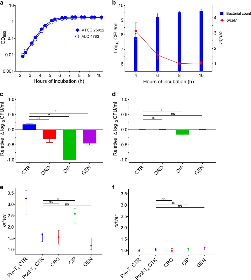
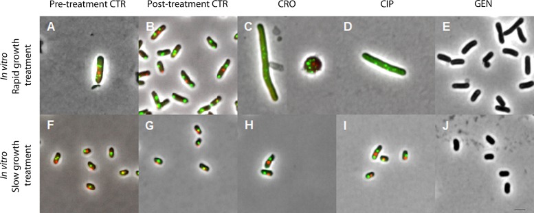
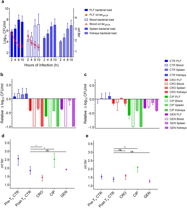
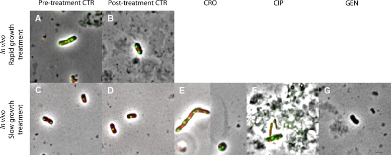
Commonly used antibiotics exert their effects predominantly on rapidly growing bacterial cells; yet, the growth dynamics taking place during infection in a complex host environment remain largely unknown. Hence, a means to measure in situ bacterial growth rate is essential to predict the outcome of antibacterial treatment. We have recently validated chromosome replication as a readout of in situ bacterial growth rate during Escherichia coli infection in the mouse peritonitis model. By the use of two complementary methods (quantitative PCR and fluorescence microscopy) for differential genome origin and terminus copy number quantification, we demonstrated the ability to track bacterial growth rate, both on a population average level and on a single-cell level, from one single biological specimen. Here, we asked whether the in situ growth rate predicts antibiotic treatment effect during infection in the same model. Parallel in vitro growth experiments were conducted as a proof of concept. Our data demonstrate that the activities of the commonly used antibiotics ceftriaxone and gentamicin correlated with pretreatment bacterial growth rate; both drugs performed better during rapid growth than during slow growth. Conversely, ciprofloxacin was less sensitive to bacterial growth rate, both in a homogenous in vitro bacterial population and in a more heterogeneous in vivo bacterial population. The method serves as a platform to test any antibiotic's dependency on active in situ bacterial growth. Improved insight into this relationship in vivo could ultimately prove helpful in evaluating future antibacterial strategies.

Keywords: Escherichia coli; bacterial growth; chromosome replication; experimental animal model.


Figures

FIG 1
FIG 1
Antibiotic activity as a function of bacterial growth rate in vitro. (a) Parallel growth of ALO 4783 relative to the ATCC 25922 wild type in LB revealed no growth retardation due to transgene insertions. Cell density measured as optical density at 600 nm (OD600). (b) Bacterial counts (CFU/ml) and growth rates (ori:ter) in untreated control batch cultures (ATCC 25922 and ALO 4783); n = 6. Data presented as means (SDs). (c) Bacterial count reductions after 2 h of antibiotic exposure in ceftriaxone (CRO), ciprofloxacin (CIP), and gentamicin (GEN) treatment batch cultures when therapy was induced during rapid bacterial growth (i.e., at 4 h of incubation). Controls (CTR) received no antibiotic therapy. (d) Bacterial count reductions after 2 h of antibiotic exposure in treatment batch cultures when therapy was induced during slow bacterial growth (i.e., at 8 h of incubation). CTR received no antibiotic therapy. For comparison of activities between treatment inductions during rapid and slow growth, data in panels c and d are presented as relative bacterial count reductions. (e) Bacterial growth rates (ori:ter) in pretreatment controls (Pre-Tx CTR) and posttreatment controls (Post-Tx CTR) (i.e., at 4 and 6 h of incubation, respectively) and in treatment batch cultures after 2 h of antibiotic exposure when therapy was induced during rapid bacterial growth. (f) Bacterial growth rates (ori:ter) in Pre-Tx and Post-Tx controls (i.e., at 8 and 10 h of incubation, respectively) and in treatment batch cultures after 2 h of antibiotic exposure when therapy was induced during slow bacterial growth. Data in panels c to f are presented as medians and interquartile ranges (IQRs). CTR, n = 6; CRO, n = 6; CIP, n = 6; GEN, n = 3. *, P < 0.05; **, P < 0.01; ns, P > 0.05 by Mann-Whitney U test.
FIG 2
FIG 2
Representative examples of pooled bacterial cells observed by fluorescence microscopy and isolated after antibiotic induction during rapid (top row) or slow (bottom row) bacterial growth in vitro. Images are shown at the same magnification (using a 100× objective) in phase contrast; intracellular oriC foci in green (green fluorescent protein [GFP]) and terC foci in red (mCherry) (ALO 4783). For GEN treatment experiments, ATCC 25922 without fluorescent foci was utilized. A total of 500 pooled cells were analyzed per time point from all cultures with the exception of rapid growth treatment cultures: CRO, n = 28; CIP, n = 112; GEN, n = 330. Due to the limited resolution of fluorescence microscopy for colocalizing oriCs, some bacterial cells with overlapping chromosome replication origins may appear with too few foci (33). Mean (SD) population medial axis cell lengths were as follows: (A) rapid bacterial growth, pretreatment CTR, 4.1 (0.98) μm; (B) rapid bacterial growth, posttreatment CTR, 3.51 (0.86) μm; (C) rapid bacterial growth, post-CRO treatment, not determined (due to overrepresentation of spherical cells); (D) rapid bacterial growth, post-CIP treatment, 7.39 (2.52) μm; (E) rapid bacterial growth, post-GEN treatment, 3.25 (0.84) μm; (F) slow bacterial growth, pretreatment CTR, 2.58 (0.66) μm; (G) slow bacterial growth, posttreatment CTR, 2.25 (0.52) μm; (H) slow bacterial growth, post-CRO treatment, 2.67 (0.67) μm; (I) slow bacterial growth, post-CIP treatment, 2.53 (0.69) μm; (J) slow bacterial growth, post-GEN treatment, 2.18 (0.45) μm. CTR, controls; CRO, ceftriaxone; CIP, ciprofloxacin; GEN, gentamicin. Scale bar, 2 μm.
FIG 3
FIG 3
Antibiotic activity as a function of bacterial growth rate during infection in vivo in the mouse peritonitis model. (a) Bacterial counts (CFU/ml; n = 9 per time point) and growth rates (ori:ter; 2, 8, and 10 h of infection, n = 9; 4 h of infection, n = 6) in untreated control groups (ATCC 25922 and ALO 4783) in the peritoneal lavage fluid (PLF), blood, spleen, and kidneys (bacterial counts only in the tissues). (b) Bacterial count reductions in PLF, blood, spleen, and kidneys after 2 h of antibiotic exposure in ceftriaxone (CRO), ciprofloxacin (CIP), and gentamicin (GEN) treatment groups when therapy was induced during rapid bacterial growth (i.e., at 2 h of infection). Controls (CTR) received no antibiotic therapy. (c) Bacterial count reductions in PLF, blood, spleen, and kidneys after 2 h of antibiotic exposure in treatment groups when therapy was induced during slow bacterial growth (i.e., at 8 h of infection). CTR received no antibiotic therapy. For comparison of activities between treatment induction during rapid and slow growth, data in panels b and c are presented as relative bacterial count reductions. CTR, n = 9; CRO, CIP, and GEN, n = 3. (d) Bacterial growth rates (ori:ter) in pretreatment controls (Pre-Tx CTR) and posttreatment controls (Post-Tx CTR) (i.e., at 2 and 4 h of infection, respectively) and in treatment groups after 2 h of antibiotic exposure when treatment was induced during rapid bacterial growth. As there was no significant difference in ori:ter values between control bacterial populations from PLF and blood, these were pooled for analysis. ori:ter values in treatment groups were only available from PLF, due to total bacterial elimination from the blood. Pretreatment CTR, n = 18; posttreatment CTR, n = 12; CRO, n = 3; CIP, n = 3; GEN, n = 3. (e) Bacterial growth rates (ori:ter) in Pre-Tx CTR and Post-Tx CTR (i.e., at 8 and 10 h of infection, respectively) and in treatment groups after 2 h of antibiotic exposure when treatment was induced during slow bacterial growth. PLF and blood ori:ter values were pooled for analysis. Pretreatment CTR, n = 18; posttreatment CTR, n = 18; CRO, n = 6; CIP, n = 3; GEN, n = 6. Data in panels b to e are presented as medians and interquartile ranges (IQRs). *, P < 0.05; **, P < 0.01; ns, P > 0.05 by Mann-Whitney U test.
FIG 4
FIG 4
Representative examples of bacterial cells observed by fluorescence microscopy and isolated after antibiotic induction during rapid (top row) or slow (bottom row) bacterial growth in vivo (blood and peritoneal lavage fluid [PLF] bacterial cells pooled). Images are shown at the same magnification (using a 100× objective) in phase contrast; intracellular oriC foci in green (GFP) and terC foci in red (mCherry) (ALO 4783). For GEN treatment experiments, ATCC 25922 without fluorescent foci was utilized. Examples shown are bacteria isolated from the PLF. Due to total or near total elimination of bacterial cells during rapid growth treatment, no bacterial cells were available from these treatment groups. A total of 500 cells were pooled and analyzed per time point from pre- and posttreatment controls during slow growth. For pre- and posttreatment controls during rapid growth, fewer cells were isolated due to low bacterial counts during early hours of infection: n = 142 and n = 66, respectively. For slow growth treatment induction: CRO, n = 170; CIP, n = 35; GEN, n = 228. Due to the limited resolution of fluorescence microscopy for colocalizing oriCs, some bacterial cells with overlapping chromosome replication origins may appear with too few foci (33). Mean (SD) population medial axis cell lengths were as follows: (A) rapid bacterial growth, pretreatment CTR, 3.96 (1.15) μm; (B) rapid bacterial growth, posttreatment CTR, 3.73 (1.13) μm; (C) slow bacterial growth, pretreatment CTR, 3.19 (0.73) μm; (D) slow bacterial growth, posttreatment CTR, 2.77 (0.86) μm; (E) slow bacterial growth, post-CRO treatment, not determined (due to overrepresentation of spherical cells); (F) slow bacterial growth, post-CIP treatment, 3.25 (0.68) μm; (G) slow bacterial growth, post-GEN treatment, 2.56 (0.76) μm. CTR, controls; CRO, ceftriaxone; CIP, ciprofloxacin; GEN, gentamicin. Scale bar, 2 μm.

References

    1. Kopf SH, Sessions AL, Cowley ES, Reyes C, Van Sambeek L, Hu Y, Orphan VJ, Kato R, Newman DK. 2016. Trace incorporation of heavy water reveals slow and heterogeneous pathogen growth rates in cystic fibrosis sputum. Proc Natl Acad Sci U S A 113:E110–E116. doi:10.1073/pnas.1512057112. - DOI - PMC - PubMed
    1. Olm MR, Brown CT, Brooks B, Firek B, Baker R, Burstein D, Soenjoyo K, Thomas BC, Morowitz M, Banfield JF. 2017. Identical bacterial populations colonize premature infant gut, skin, and oral microbiomes and exhibit different in situ growth rates. Genome Res 27:601–612. doi:10.1101/gr.213256.116. - DOI - PMC - PubMed
    1. Haugan MS, Charbon G, Frimodt-Møller N, Løbner-Olesen A. 2018. Chromosome replication as a measure of bacterial growth rate during Escherichia coli infection in the mouse peritonitis model. Sci Rep 8:14961. doi:10.1038/s41598-018-33264-7. - DOI - PMC - PubMed
    1. Lee AJ, Wang S, Meredith HR, Zhuang B, Dai Z, You L. 2018. Robust, linear correlations between growth rates and β-lactam-mediated lysis rates. Proc Natl Acad Sci U S A 115:4069–4074. doi:10.1073/pnas.1719504115. - DOI - PMC - PubMed
    1. Tuomanen E, Cozens R, Tosch W, Zak O, Tomasz A. 1986. The rate of killing of Escherichia coli by β-lactam antibiotics is strictly proportional to the rate of bacterial growth. J Gen Microbiol 132:1297–1304. doi:10.1099/00221287-132-5-1297. - DOI - PubMed

Publication types

MeSH terms