. 2018 Dec 18;115(51):13117-13122.
doi: 10.1073/pnas.1812282115. Epub 2018 Dec 3.

Constitutive boost of a K+ channel via inherent bilayer tension and a unique tension-dependent modality

Affiliations

Constitutive boost of a K+ channel via inherent bilayer tension and a unique tension-dependent modality

Masayuki Iwamoto et al. Proc Natl Acad Sci U S A. .

Abstract

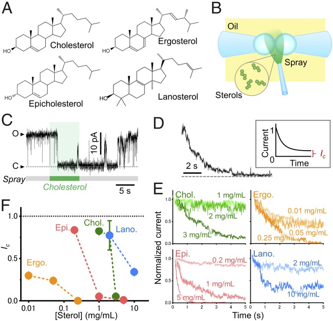
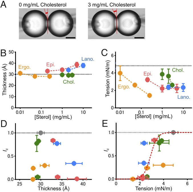
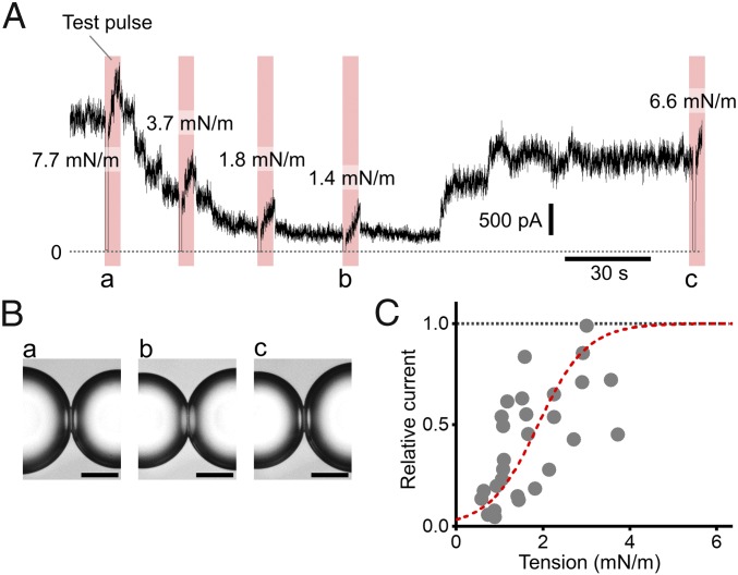
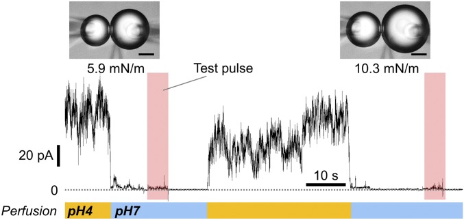
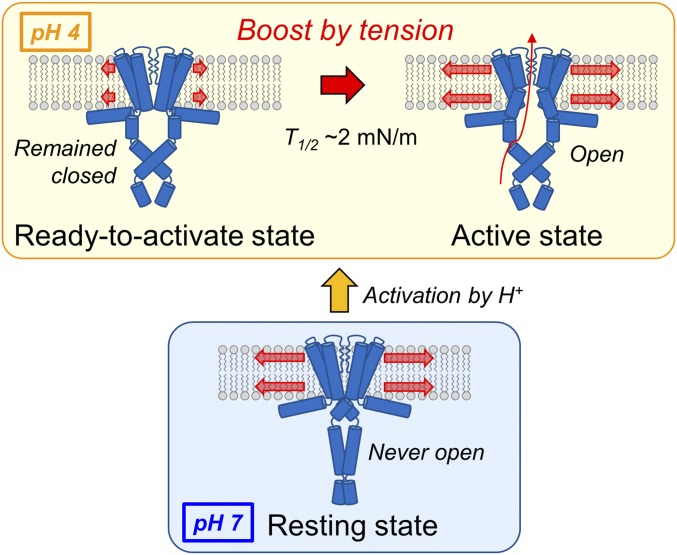
Molecular mechanisms underlying channel-membrane interplay have been extensively studied. Cholesterol, as a major component of the cell membrane, participates either in specific binding to channels or via modification of membrane physical features. Here, we examined the action of various sterols (cholesterol, epicholesterol, etc.) on a prototypical potassium channel (KcsA). Single-channel current recordings of the KcsA channel were performed in a water-in-oil droplet bilayer (contact bubble bilayer) with a mixed phospholipid composition (azolectin). Upon membrane perfusion of sterols, the activated gate at acidic pH closed immediately, irrespective of the sterol species. During perfusion, we found that the contacting bubbles changed their shapes, indicating alterations in membrane physical features. Absolute bilayer tension was measured according to the principle of surface chemistry, and inherent bilayer tension was ∼5 mN/m. All tested sterols decreased the tension, and the nonspecific sterol action to the channel was likely mediated by the bilayer tension. Purely mechanical manipulation that reduced bilayer tension also closed the gate, whereas the resting channel at neutral pH never activated upon increased tension. Thus, rather than conventional stretch activation, the channel, once ready to activate by acidic pH, changes the open probability through the action of bilayer tension. This constitutes a channel regulating modality by two successive stimuli. In the contact bubble bilayer, inherent bilayer tension was high, and the channel remained boosted. In the cell membrane, resting tension is low, and it is anticipated that the ready-to-activate channel remains closed until bilayer tension reaches a few millinewton/meter during physiological and pathological cellular activities.

Keywords: KcsA channel; bilayer tension; contact bubble bilayer; single-channel current; stretch-activated channel.


Figures

Fig. 1.
Fig. 1.
Effect of sterol administration to the membrane on the KcsA channel current. (A) Chemical structures of sterols used in this paper. (B) Schematic of the membrane perfusion of sterols. Sterols dissolved in hexadecane were sprayed around the CBB and were immediately transferred into the membrane interior. (C) A typical single-channel current response to cholesterol perfusion. (D) An ensemble averaged trace upon perfusion of 3 mg/mL cholesterol with time 0 set at the onset of perfusion. (Inset) The residual current amplitude was obtained from the ensemble averaged current traces, and this current amplitude relative to that before cholesterol perfusion was defined as Ic. (E) Ensemble averaged traces upon perfusion of various sterols. Each sterol was dissolved in hexadecane at the desired concentration and perfused at time 0. The number of raw traces for the ensemble average was 5–24. (F) Sterol concentration dependencies of Ic. The error bar indicates the standard error of the mean (SEM) for the regression (n = 3–6).
Fig. 2.
Fig. 2.
Effect of sterols on bilayer tension and thickness. (A) Shape changes in the CBB in the absence or presence (3 mg/mL) of cholesterol. The bilayer area expanded, and the contact angle increased. The bar represents 50 μm. (B) Bilayer thickness as a function of sterol concentration. The dotted black line indicates thickness in the absence of sterol. (C) Bilayer tension as a function of sterol concentration. The dotted black line indicates bilayer tension in the absence of sterol. (D and E) Relationship between Ic values and bilayer thickness (D) or tension (E). The broken red line in (E) represents a Boltzmann fit (see Discussion). Cholesterol (green), epicholesterol (red), ergosterol (orange), and lanosterol (blue). The error bars indicate the SEM (n = 3–8).
Fig. 3.
Fig. 3.
Manipulation of lipid bilayer tension and response of the KcsA channel. (A) Macroscopic KcsA current responses to changes in bilayer tension. Lipid bilayer tension was manipulated by controlling the bubble-maintaining pressure of both bubbles. Initial bilayer tension was set high (>4 mN/m), and macroscopic current was measured at +100 mV. The reduction in tension attenuated the current amplitude, so it reached a steady level where the contact angle was evaluated at different membrane potentials (magenta belt; SI Appendix, Fig. S2A). (B) Shape changes upon alterations in bubble pressure at the instance denoted as ac in A. The bar represents 50 μm. (C) Bilayer tension dependency of the KcsA channel activity. Relative current amplitudes (I/Ihigh tension) are plotted as a function of bilayer tension. The broken red line represents a Boltzmann fit where with ΔG0/kBT of 3.37 ± 0.37, ΔA of 7.33 ± 1.23 nm2, and T1/2 of 1.89 ± 0.14 nM/m.
Fig. 4.
Fig. 4.
Stretch activation of the resting KcsA channel at neutral pH. Two micropipettes that enabled changes in pH were inserted into the right bubble. The macroscopic channel current recorded at acidic pH was attenuated as the cytosolic side was perfused to neutral pH. Then, bilayer tension was applied by pressurizing both bubbles during which the test pulse was applied and bilayer tension was evaluated (magenta belt). The current failed to be elicited by increased bilayer tension even up to 10.3 mN/m. As the cytosolic pH was returned to acidic, the channel current was elicited.
Fig. 5.
Fig. 5.
Schematic of the KcsA channel responding to bilayer tension. When the channel remained in the resting state at neutral intracellular pH, it never opened upon increased bilayer tension. At acidic pH, the channel exhibited variable activity depending on bilayer tension (tension boosting). However, the channel stayed closed below a bilayer tension of 2 mN/m.

Similar articles

Cited by

References

    1. Tillman TS, Cascio M. Effects of membrane lipids on ion channel structure and function. Cell Biochem Biophys. 2003;38:161–190. - PubMed
    1. Dufourc EJ. Sterols and membrane dynamics. J Chem Biol. 2008;1:63–77. - PMC - PubMed
    1. Nicolson GL. The fluid-mosaic model of membrane structure: Still relevant to understanding the structure, function and dynamics of biological membranes after more than 40 years. Biochim Biophys Acta. 2014;1838:1451–1466. - PubMed
    1. Simons K, Sampaio JL. Membrane organization and lipid rafts. Cold Spring Harb Perspect Biol. 2011;3:a004697. - PMC - PubMed
    1. Levitan I, Singh DK, Rosenhouse-Dantsker A. Cholesterol binding to ion channels. Front Physiol. 2014;5:65. - PMC - PubMed

Publication types