. 2018 Dec 18;115(51):E12053-E12062.
doi: 10.1073/pnas.1813365115. Epub 2018 Dec 3.

Synergistic neuroprotection by coffee components eicosanoyl-5-hydroxytryptamide and caffeine in models of Parkinson's disease and DLB

Affiliations

Synergistic neuroprotection by coffee components eicosanoyl-5-hydroxytryptamide and caffeine in models of Parkinson's disease and DLB

Run Yan et al. Proc Natl Acad Sci U S A. .

Abstract

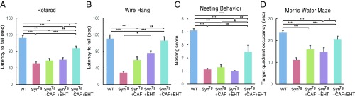
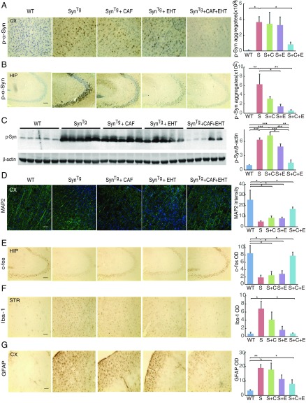
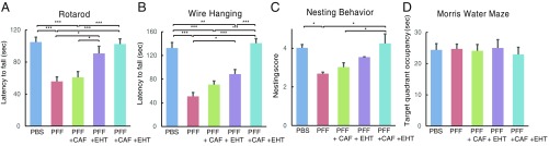
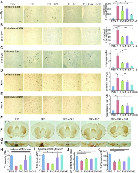
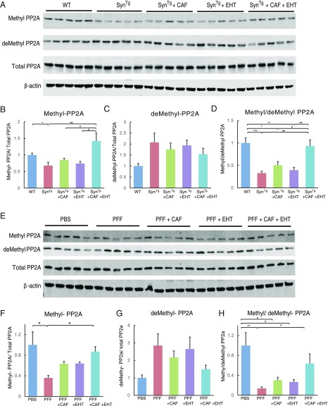
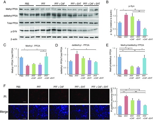
Hyperphosphorylated α-synuclein in Lewy bodies and Lewy neurites is a characteristic neuropathological feature of Parkinson's disease (PD) and Dementia with Lewy bodies (DLB). The catalytic subunit of the specific phosphatase, protein phosphatase 2A (PP2A) that dephosphorylates α-synuclein, is hypomethylated in these brains, thereby impeding the assembly of the active trimeric holoenzyme and reducing phosphatase activity. This phosphatase deficiency contributes to the accumulation of hyperphosphorylated α-synuclein, which tends to fibrillize more than unmodified α-synuclein. Eicosanoyl-5-hydroxytryptamide (EHT), a fatty acid derivative of serotonin found in coffee, inhibits the PP2A methylesterase so as to maintain PP2A in a highly active methylated state and mitigates the phenotype of α-synuclein transgenic (SynTg) mice. Considering epidemiologic and experimental evidence suggesting protective effects of caffeine in PD, we sought, in the present study, to test whether there is synergy between EHT and caffeine in models of α-synucleinopathy. Coadministration of these two compounds orally for 6 mo at doses that were individually ineffective in SynTg mice and in a striatal α-synuclein preformed fibril inoculation model resulted in reduced accumulation of phosphorylated α-synuclein, preserved neuronal integrity and function, diminished neuroinflammation, and improved behavioral performance. These indices were associated with increased levels of methylated PP2A in brain tissue. A similar profile of greater PP2A methylation and cytoprotection was found in SH-SY5Y cells cotreated with EHT and caffeine, but not with each compound alone. These findings suggest that these two components of coffee have synergistic effects in protecting the brain against α-synuclein-mediated toxicity through maintenance of PP2A in an active state.

Keywords: Dementia with Lewy bodies; Parkinson’s disease; neuroprotection; phosphorylation; α-synuclein.


Conflict of interest statement

Conflict of interest statement: J.B.S. has a financial interest in Signum Biosciences, which is developing PP2A phosphatase enhancing agents. M.V. is employed by Signum Biosciences. J.B.S. and M.M.M. are inventors of a patent application relevant to this work.

Figures

Fig. 1.
Fig. 1.
EHT and CAF cotreatment prevents the behavioral deficits of SynTg mice. Behavioral performance of WT and SynTg mice (WT, n = 23; SynTg, n = 20; SynTg+CAF, n = 16; SynTg+EHT, n = 14; SynTg+CAF+EHT, n = 21) were tested at 6 mo of age on the (A) rotarod, (B) Wire Hang, (C) nesting behavior, and (D) Morris Water Maze. Data shown represent means ± SEM. *P < 0.05; **P < 0.01; ***P < 0.001.
Fig. 2.
Fig. 2.
EHT and CAF cotreatment reduces p-α-Syn burden and protects against the neuronal damage and neuroinflammation in SynTg mice. (A and B) Representative images and quantification of immunohistochemical staining of p-α-Syn in the (A) cortex and (B) hippocampus. (C) Western blot analysis for p-α-Syn and β-actin with cortical brain tissue lysates from five groups and five animals per group. (D) Representative images and quantification of immunofluorescence staining of MAP2 in the cortex. (E) Representative images and quantification of immunohistochemical staining of c-fos in the hippocampus. (F) Representative images and quantification of immunohistochemical staining of Iba-1 in the striatum. (G) Representative images and quantification of immunohistochemical staining of GFAP in the cortex. (For all images, scale bar: 100 μm.) All bar graphs represent means ± SEM; n = 5 to 6 mice for immunohistochemical and immunofluorescence stains. *P < 0.05; **P < 0.01; ***P < 0.001.
Fig. 3.
Fig. 3.
EHT and CAF cotreatment improves the behavioral performance of α-Syn PFF-inoculated WT mice. Behavioral performance of PBS- and α-Syn PFF-inoculated mice (PBS, n = 14; PFF, n = 14; PFF+CAF, n = 19; PFF+EHT, n = 14; PFF+CAF+EHT, n = 15) was tested 6 mo postinoculation, at 8 mo of age on the (A) rotarod, (B) Wire Hang, (C) nesting behavior, and (D) Morris Water Maze. Bar graphs represent means ± SEM. *P < 0.05; **P < 0.01; ***P < 0.001.
Fig. 4.
Fig. 4.
EHT and CAF cotreatment prevents the formation of p-α-Syn−positive aggregates, mitigates neuroinflammation, and protects nigrostriatal neurons in the α-Syn PFF inoculation model. (A and B) Representative images and quantification of immunohistochemical staining of p-α-Syn in the (A) ipsilateral striatum and (B) contralateral striatum. (C) Representative images and quantification of immunohistochemical staining of p-α-Syn in the ipsilateral substantia nigra pars compacta (SNc). In AC, n = 4 to 6 per group. (D and E) Representative images and quantification of immunohistochemical staining of Iba-1 in the (D) ipsilateral and (E) contralateral striatum. In D and E, n = 5 per group. (Scale bar: 100 μm for AE.) (F) Representative images of immunohistochemical staining of TH in the striatum. (G) Representative images of immunohistochemical staining of TH in the SNc. (H and I) Quantification of (H) ipsilateral and (I) contralateral striatal TH staining; n = 5 per group. (J) DA content in the ipsilateral striatum analyzed by HPLC-MS; n = 5 to 6 per group. (K) Nigral TH-positive neuron count; n = 4 to 5 per group. All bar graphs represent means ± SEM. *P < 0.05; **P < 0.01; ***P < 0.001. C, CAF; E, EHT; F, PFF.
Fig. 5.
Fig. 5.
EHT and CAF exert their synergistic neuroprotective effects through regulating PP2A activity. (A) Western blots and (BD) densitometric analysis for the indicated proteins with striatal tissue lysates from SynTg mouse brains. Bar graphs show (B) methyl-PP2A and (C) demethyl-PP2A levels that are normalized to total PP2A, and (D) the ratio of methylated PP2A over demethyl-PP2A. (E) Western blot and (FH) densitometric analysis of ipsilateral striatal tissue lysates from α-Syn PFF-inoculated mice probed for the indicated proteins. Bar graphs show (F) methyl-PP2A and (G) demethyl-PP2A levels that are normalized to total PP2A, and (H) the ratio of methyl-PP2A over demethyl-PP2A. For all images, n = 5 per group. All bar graphs represent means ± SEM. *P < 0.05; **P < 0.01; ***P < 0.001.
Fig. 6.
Fig. 6.
Combination of EHT and CAF has synergistic effects in up-regulating PP2A methylation and attenuating cytotoxicity induced by α-Syn PFF in SH-SY5Y cells. (AE) SH-SY5Y cells were incubated with PBS or mouse α-Syn PFF and treated with CAF and/or EHT for 7 d. Cell lysates were subjected to Western blot analysis for the indicated proteins. The experiment was done in triplicates and repeated at least three times, yielding similar results. All lanes in the top Western blot (methyl-PP2A) were run on one gel. The vertical space between lanes 13 and 14 denotes where a size marker was loaded and was subsequently spliced out. (F) Representative images of PI and DAPI staining of SH-SY5Y cells incubated with PBS or mouse α-Syn PFF and treated with CAF and/or EHT for 7 d. Five random fields per well, and four independent wells per group, were counted for PI-positive cells. PI-positive cell numbers were normalized to DAPI count in the same field. Experiments were repeated twice, yielding similar results. (Scale bar: 100 μm.) All bar graphs represent means ± SEM. *P < 0.05; **P < 0.01; ***P < 0.001.

References

    1. Spillantini MG, Crowther RA, Jakes R, Hasegawa M, Goedert M. Alpha-synuclein in filamentous inclusions of Lewy bodies from Parkinson’s disease and dementia with Lewy bodies. Proc Natl Acad Sci USA. 1998;95:6469–6473. - PMC - PubMed
    1. Goedert M. Alpha-synuclein and neurodegenerative diseases. Nat Rev Neurosci. 2001;2:492–501. - PubMed
    1. Luk KC, et al. Pathological α-synuclein transmission initiates Parkinson-like neurodegeneration in nontransgenic mice. Science. 2012;338:949–953. - PMC - PubMed
    1. Braak H, Del Tredici K. Neuropathological staging of brain pathology in sporadic Parkinson’s disease: Separating the wheat from the chaff. J Parkinsons Dis. 2017;7(Suppl 1):S71–S85. - PMC - PubMed
    1. Fujiwara H, et al. Alpha-synuclein is phosphorylated in synucleinopathy lesions. Nat Cell Biol. 2002;4:160–164. - PubMed

Publication types

MeSH terms