. 2019 Jan 8;116(2):575-580.
doi: 10.1073/pnas.1810200115. Epub 2018 Dec 3.

Autocrine regulation of mesenchymal progenitor cell fates orchestrates tooth eruption

Affiliations

Autocrine regulation of mesenchymal progenitor cell fates orchestrates tooth eruption

Akira Takahashi et al. Proc Natl Acad Sci U S A. .

Abstract

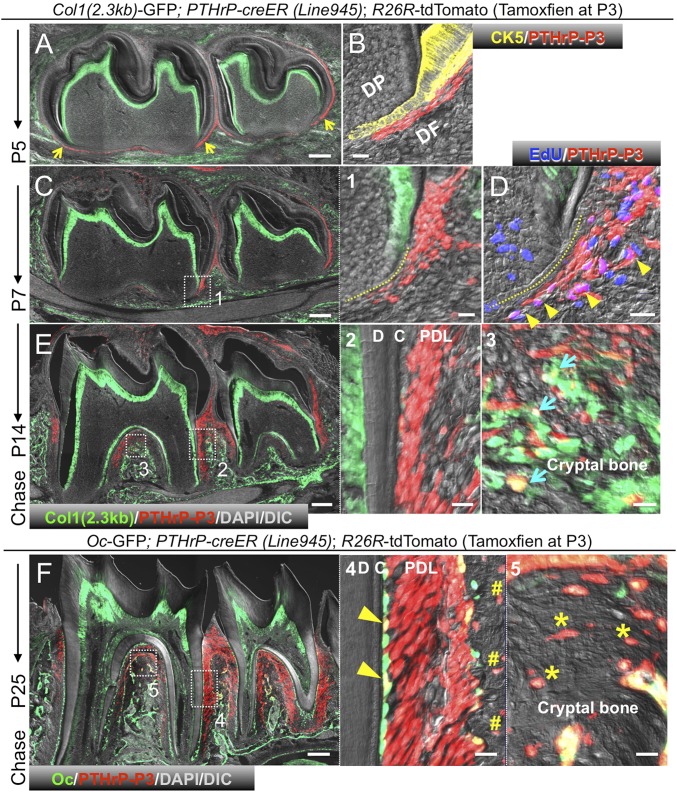
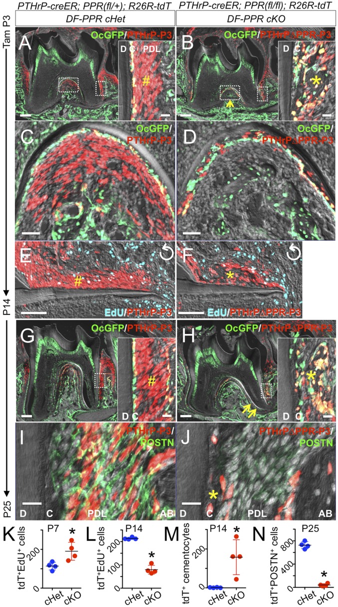
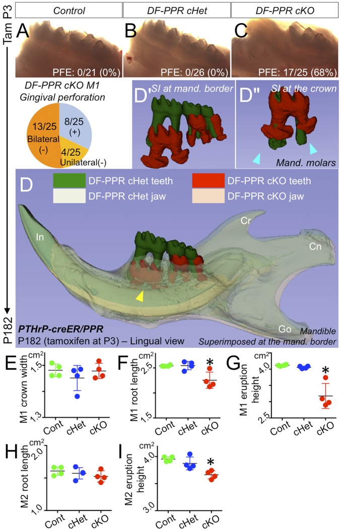
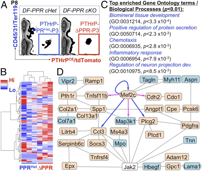
Formation of functional skeletal tissues requires highly organized steps of mesenchymal progenitor cell differentiation. The dental follicle (DF) surrounding the developing tooth harbors mesenchymal progenitor cells for various differentiated cells constituting the tooth root-bone interface and coordinates tooth eruption in a manner dependent on signaling by parathyroid hormone-related peptide (PTHrP) and the PTH/PTHrP receptor (PPR). However, the identity of mesenchymal progenitor cells in the DF and how they are regulated by PTHrP-PPR signaling remain unknown. Here, we show that the PTHrP-PPR autocrine signal maintains physiological cell fates of DF mesenchymal progenitor cells to establish the functional periodontal attachment apparatus and orchestrates tooth eruption. A single-cell RNA-seq analysis revealed cellular heterogeneity of PTHrP+ cells, wherein PTHrP+ DF subpopulations abundantly express PPR. Cell lineage analysis using tamoxifen-inducible PTHrP-creER mice revealed that PTHrP+ DF cells differentiate into cementoblasts on the acellular cementum, periodontal ligament cells, and alveolar cryptal bone osteoblasts during tooth root formation. PPR deficiency induced a cell fate shift of PTHrP+ DF mesenchymal progenitor cells to nonphysiological cementoblast-like cells precociously forming the cellular cementum on the root surface associated with up-regulation of Mef2c and matrix proteins, resulting in loss of the proper periodontal attachment apparatus and primary failure of tooth eruption, closely resembling human genetic conditions caused by PPR mutations. These findings reveal a unique mechanism whereby proper cell fates of mesenchymal progenitor cells are tightly maintained by an autocrine system mediated by PTHrP-PPR signaling to achieve functional formation of skeletal tissues.

Keywords: dental follicle; in vivo lineage-tracing experiment; mesenchymal progenitor cells; parathyroid hormone-related peptide; tooth eruption.


Conflict of interest statement

The authors declare no conflict of interest.

Figures

Fig. 1.
Fig. 1.
Characterization of PTHrP-mCherry+ DF cells. (A and B) PTHrP-mCherry expression in PTHrP+/+ and PTHrPmCherry/+ DF cells at P5. (C and D) t-SNE–based visualization of major classes of FACS-sorted PTHrP-mCherry+ single cells at P6 (clusters 0–11). Cluster 0, transitional cells; cluster 1, DF cells; cluster 2, epithelial cells; cluster 3, cycling cells; clusters 4, 6, and 10, odontoblasts and dental papilla (DP) cells; clusters 5, 8, and 11, impurified cells; clusters 7 and 9, fibroblasts. Dots indicate individual cells; color indicates cell type. (D) Pthlh and Pth1r expression. Blue, high expression; gray, no expression; red contour, DF cells. (E) MAGIC imputation analysis showing the Pthlh-Pth1r relationship (DF cells). Red arrows indicate PthlhhighPth1r+ cells.
Fig. 2.
Fig. 2.
PTHrP-creER marks DF mesenchymal progenitor cells in vivo. (AF) Lineage tracing of P3 PTHrP-creER+ DF cells. Yellow arrowheads in A indicate tdTomato+ DF cells in periapical areas. (B) Cytokeratin 5 (CK5) staining. (C) Yellow dotted line in (1) denotes HERS. (D) EdU was administered shortly before analysis. Arrowheads in D denote EdU+tdTomato+ DF cells. (E) Blue arrows in (3) indicate Col1-GFP+tdTomato+ osteoblasts in cryptal bones. (F) Yellow arrowheads in (4) indicate GFP+tdTomato+ cementoblasts on root surface. Pound signs in (4) denote tdTomato+ osteocytes in interseptic cryptal bones. Asterisks in (5) indicate tdTomato+ osteocytes in interradicular cryptal bones. C, cementum; D, dentin. PDL, periodontal ligament. [Scale bars: 200 µm (Left), 20 µm (Right).]
Fig. 3.
Fig. 3.
PTHrP-PPR autocrine signaling orchestrates PDL differentiation of DF mesenchymal progenitor cells. Conditional deletion of PPR in PTHrP-creER+ DF cells. (AF) P14. (Insets) Distal PDL space. The pound sign in A indicates tdTomato+ PDL cells. In B, the asterisk denotes the precocious cellular cementum-like structure, and the arrow denotes underdeveloped cryptal bones. In E and F, EdU was administered shortly before analysis. Perpendicularly rotated images. (GJ) P25. The pound sign in G indicates tdTomato+ PDL cells. In H, the asterisk denotes the cellular cementum-like structure, and the arrows indicate root dilacerations. (I and J) POSTN staining. The asterisk in J indicates the cellular cementum-like structure. C, cementum; D, dentin. (Scale bars: 200 µm, except 20 µm in Insets and I and J, 50 µm in C and D, and 100 µm in E and F.) (KN) Quantification of tdTomato+EdU+ cells and tdTomato+ cells per 200-µm thickness. *P < 0.05, Mann–Whitney U test. All data are mean ± SD.
Fig. 4.
Fig. 4.
PPR deletion in PTHrP-creER+ DF cells recapitulates human primary failure of eruption conditions. (AC) PFE phenotypes at 6 mo. (Pie chart) Incidence of failure of eruption (lack of gingival perforation). (D) 3D micro CT surface model overlays: superimposition of registered DF-PPR cHet and DF-PPR cKO mandibles. The yellow arrowhead indicates the first molar associated with pronounced PFE phenotypes and tooth root anomalies. Cn, condyle; Cr, coronoid process; Go, gonial angle; In, incisor. (D′ and D″) Composite 3D surface model overlay of the mandibular first molars. Blue arrowheads indicate truncation (short roots) associated with dilacerations (curved roots). (FI) Quantitative 3D micro CT analysis. *P < 0.05, one-way ANOVA followed by the Mann–Whitney U test. All data are mean ± SD.
Fig. 5.
Fig. 5.
RNA-seq analysis of PPR-deficient PTHrP-creER+ DF mesenchymal progenitor cells. (A) FACS-sorting of PTHrP-creER+ DF cells. Blue box, PTHrP-PPRHet-P3 cells (cHet); red box, PTHrP-∆PPR-P3 cells (cKO). (BD) RNA-seq analysis of DF cells. (B) Heatmap of 122 DEGs with hierarchical clustering. Red indicates higher expression; blue, lower expression. (C) GO terms/biological processes overrepresented in DEGs (P[elim] < 0.01; minimum number of DEGs, 6). (D) STRING, Cytoscape, and Genomatix network and transcription factor analysis of DEGs. Oval denotes transcription factor; hexagons, matrix genes. Beige indicates up-regulated genes; blue, down-regulated genes. Blue arrows indicate validated targets; magenta arrows, predicted targets.

Comment in

Similar articles

Cited by

References

    1. Ono N, Kronenberg HM. Bone repair and stem cells. Curr Opin Genet Dev. 2016;40:103–107. - PMC - PubMed
    1. Sacchetti B, et al. Self-renewing osteoprogenitors in bone marrow sinusoids can organize a hematopoietic microenvironment. Cell. 2007;131:324–336. - PubMed
    1. Mizuhashi K, et al. Resting zone of the growth plate houses a unique class of skeletal stem cells. Nature. 2018;563:254–258. - PMC - PubMed
    1. Zhao H, et al. The suture provides a niche for mesenchymal stem cells of craniofacial bones. Nat Cell Biol. 2015;17:386–396. - PMC - PubMed
    1. Philbrick WM, Dreyer BE, Nakchbandi IA, Karaplis AC. Parathyroid hormone-related protein is required for tooth eruption. Proc Natl Acad Sci USA. 1998;95:11846–11851. - PMC - PubMed

Publication types

MeSH terms

Substances