. 2019 Jan 1;202(1):292-299.
doi: 10.4049/jimmunol.1800878. Epub 2018 Dec 3.

Improved Multiplex Immunohistochemistry for Immune Microenvironment Evaluation of Mouse Formalin-Fixed, Paraffin-Embedded Tissues

Affiliations

Improved Multiplex Immunohistochemistry for Immune Microenvironment Evaluation of Mouse Formalin-Fixed, Paraffin-Embedded Tissues

Noah Sorrelle et al. J Immunol. .

Abstract

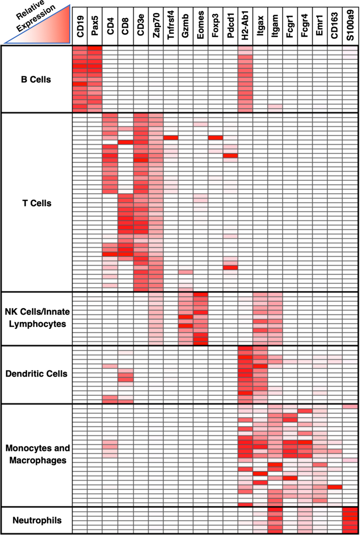
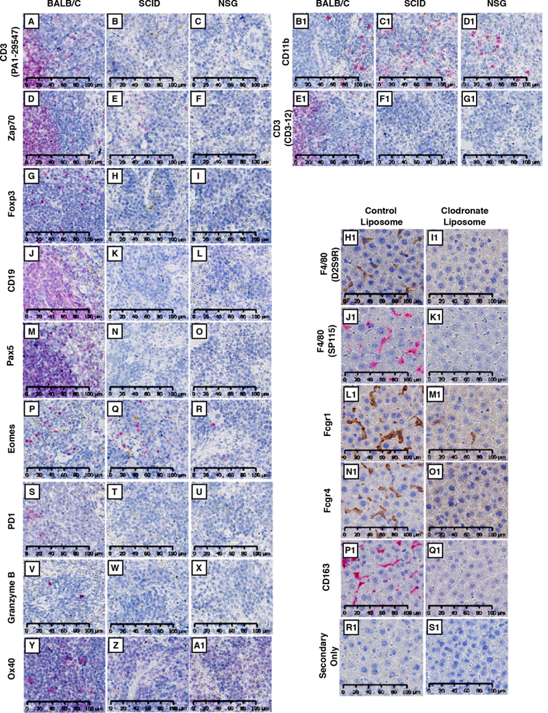
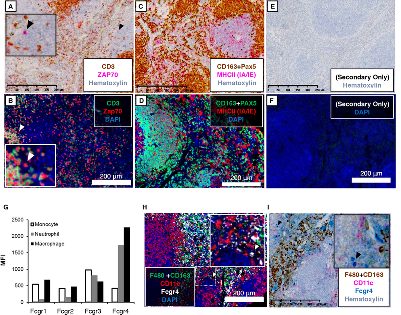
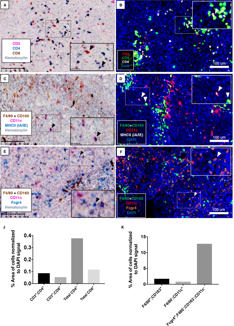
Immune profiling of tissue through multiplex immunohistochemistry is important for the investigation of immune cell dynamics, and it can contribute to disease prognosis and evaluation of treatment response in cancer patients. However, protocols for mouse formalin-fixed, paraffin-embedded tissue have been less successful. Given that formalin fixation and paraffin embedding remains the most common preparation method for processing mouse tissue, this has limited the options to study the immune system and the impact of novel therapeutics in preclinical models. In an attempt to address this, we developed an improved immunohistochemistry protocol with a more effective Ag-retrieval buffer. We also validated 22 Abs specific for mouse immune cell markers to distinguish B cells, T cells, NK cells, macrophages, dendritic cells, and neutrophils. In addition, we designed and tested novel strategies to identify immune cells for which unique Abs are currently not available. Last, in the 4T1 model of breast cancer, we demonstrate the utility of our protocol and Ab panels in the quantitation and spatial distribution of immune cells.


Conflict of interest statement

Conflict of Interests: none

Figures

Figure 1.
Figure 1.. Heatmap depicting immune cell expression of markers utilized in our immunohistochemistry panel.
Micro-array data from ImmGen showing relative expression levels of different markers by different cell types (http://www.immgen.org ) (20). Rows represent different immune cell populations, grouped by cell type. Columns represent genes.
Figure 2.
Figure 2.. Validation of antibody specificity.
(A-G1) Wild type BALB/c, SCID, or NSG spleens were probed with antibodies specific for CD3 (PA1–29547; A, B, C), Zap70 (D, E, F), Foxp3 (G, H, I), CD19 (J, K, L), Pax5 (M, N, O), Eomes (P, Q, R), PD1(S, T, U), Granzyme B (V, W, X), Ox40 (Y, Z, A1), CD11b (B1, C1, D1), and CD3 (CD3–12; E1, F1, G1). For detection, Warp Red chromogenic substrate was used. Slides were counterstained with hematoxylin and scanned at 20X using Hamamatsu NanoZoomer 2.0-HT (H1-S1) Livers from control- or clodronate-liposome treated BALB/c mice were stained for F4/80 (D2S9R and SP115; H1, I1 and J1, K1, respectively), Fcgr1 (L1, M1), Fcgr4 (N1, O1), CD163 (P1, Q1), or secondary-only (R1, S1). Slides were scanned at 20X using the Hamamatsu NanoZoomer 2.0-HT. All images represent a 40X field-of-view. Scale bar = 100 μm for all images.
Figure 3.
Figure 3.. Multiplex IHC to stain Natural Killer Cells, Dendritic cells and Neutrophils.
BALB/c spleens were stained for (A, B) CD3 (PA1–29547, T cells) and Zap70 (T cells, NK cells); (C, D) Pax5 (B cells), CD163 (majorly macrophages), and MHCII (IA/IE); (H, I) F4/80 (SP115), CD163, CD11c, and Fcgr4. (E, F) Secondary-antibody-only (without addition of primary antibody) was used for negative controls. Chromogenic substrates used were Betazoid DAB (brown) and Warp Red (pink). Opal 520 (green), Opal 570 (red) and Opal 690 (white) were used for fluorescence detection. Chromogen-stained sections were counterstained with hematoxylin. For fluorescence detection, sections were counterstained with DAPI (blue). Slides were scanned at 20X using the Hamamatsu NanoZoomer 2.0-HT (chromagen detection) or the Zeiss Axioscan.Z1 (fluorescence detection). Images represent a 10x field-of-view (G) Total splenocytes were isolated from C57BL/6 mice. Fcgr expression was evaluated on neutrophils (CD11b+; Ly6G+; Ly6C-), macrophages (F4/80+), and monocytes (CD11b+; Ly6c+Ly6G-). Fcgr signal is represented by mean fluorescence intensity. Scale bar = 250 μm for chromagen-stained images. Scale bar = 200 μm for fluorescent images.
Figure 4.
Figure 4.. Multiplex IHC staining of 4T1 tumor tissues.
4T1 primary tumors were stained for (A, B) CD3 (PA1–29547; T cells), CD4 (EPR19514; T cells, NK cells, DCs), and CD8 (T cells, NK cells, DCs); (C, D), CD11c, Fcgr4, and CD163+F4/80(SP115); (E, F) CD11c, MHCII (IA/IE), and CD163+F4/80(SP115). Chromogenic substrates used were Betazoid DAB (brown), Warp Red (pink), and Feranji Blue (blue) (A, C, E). Opal 520 (green), Opal 570 (red), and Opal 690 (white or orange) were used for fluorescent staining (B, D, F). Chromogen sections were counterstained with hematoxylin, whereas DAPI staining was used for fluorescence detetction. Slides were scanned at 20X using the Hamamatsu NanoZoomer 2.0-HT (chromagen detection) or the Zeiss Axioscan.Z1 (fluorescence detection). Images represent a 20x field-of-view. Inset: Higher magnification. (B, C, D) Arrows point to CD3+;CD8+ (B), CD11c+;MHCII+ (C; D, left arrow), or CD163&F4/80+;CD11c+ cells (D, right arrow). (E, F) Arrows point to Fcgr4+; CD11c- CD163&F4/80- cells. (J, K) Cell types in 4T1 tumor sections were quantitated as % area of positive cells normalized to DAPI signal. Scale bar = 100 μm for all images.

References

    1. Szantho E, Karai B, Ivady G, Bedekovics J, Szegedi I, Petras M, Ujj G, Ujfalusi A, Kiss C, Kappelmayer J, and Hevessy Z 2018. Comparative Analysis of Multicolor Flow Cytometry and Immunohistochemistry for the Detection of Disseminated Tumor Cells. Appl Immunohistochem Mol Morphol 26: 305–315. - PubMed
    1. Saravanan L, and Juneja S 2010. Immunohistochemistry is a more sensitive marker for the detection of myeloperoxidase in acute myeloid leukemia compared with flow cytometry and cytochemistry. Int J Lab Hematol 32: e132–136. - PubMed
    1. Koyama S, Akbay EA, Li YY, Aref AR, Skoulidis F, Herter-Sprie GS, Buczkowski KA, Liu Y, Awad MM, Denning WL, Diao L, Wang J, Parra-Cuentas ER, Wistuba II, Soucheray M, Thai T, Asahina H, Kitajima S, Altabef A, Cavanaugh JD, Rhee K, Gao P, Zhang H, Fecci PE, Shimamura T, Hellmann MD, Heymach JV, Hodi FS, Freeman GJ, Barbie DA, Dranoff G, Hammerman PS, and Wong KK 2016. STK11/LKB1 Deficiency Promotes Neutrophil Recruitment and Proinflammatory Cytokine Production to Suppress T-cell Activity in the Lung Tumor Microenvironment. Cancer Res 76: 999–1008. - PMC - PubMed
    1. Sridharan V, Gjini E, Liao X, Chau NG, Haddad RI, Severgnini M, Hammerman P, El-Naggar A, Freeman GJ, Hodi FS, Rodig SJ, Dranoff G, and Schoenfeld JD 2016. Immune Profiling of Adenoid Cystic Carcinoma: PD-L2 Expression and Associations with Tumor-Infiltrating Lymphocytes. Cancer Immunol Res 4: 679–687. - PubMed
    1. Ludwig KF, Du W, Sorrelle NB, Wnuk-Lipinska K, Topalovski M, Toombs JE, Cruz VH, Yabuuchi S, Rajeshkumar NV, Maitra A, Lorens JB, and Brekken RA 2018. Small-Molecule Inhibition of Axl Targets Tumor Immune Suppression and Enhances Chemotherapy in Pancreatic Cancer. Cancer Res 78: 246–255. - PMC - PubMed

Publication types

MeSH terms