. 2018 Dec 3;8(1):17531.
doi: 10.1038/s41598-018-35719-3.

Impaired male fertility and abnormal epididymal epithelium differentiation in mice lacking CRISP1 and CRISP4

Affiliations

Impaired male fertility and abnormal epididymal epithelium differentiation in mice lacking CRISP1 and CRISP4

Guillermo Carvajal et al. Sci Rep. .

Abstract

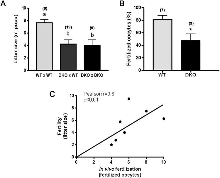
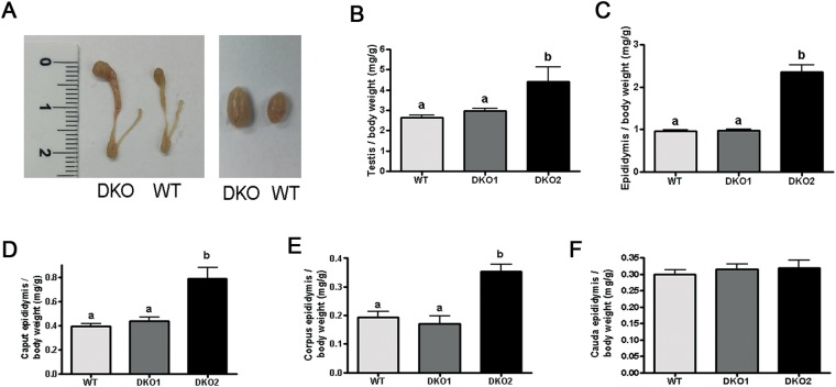
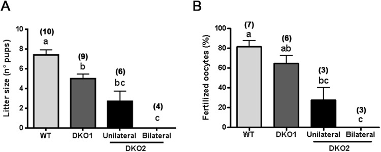
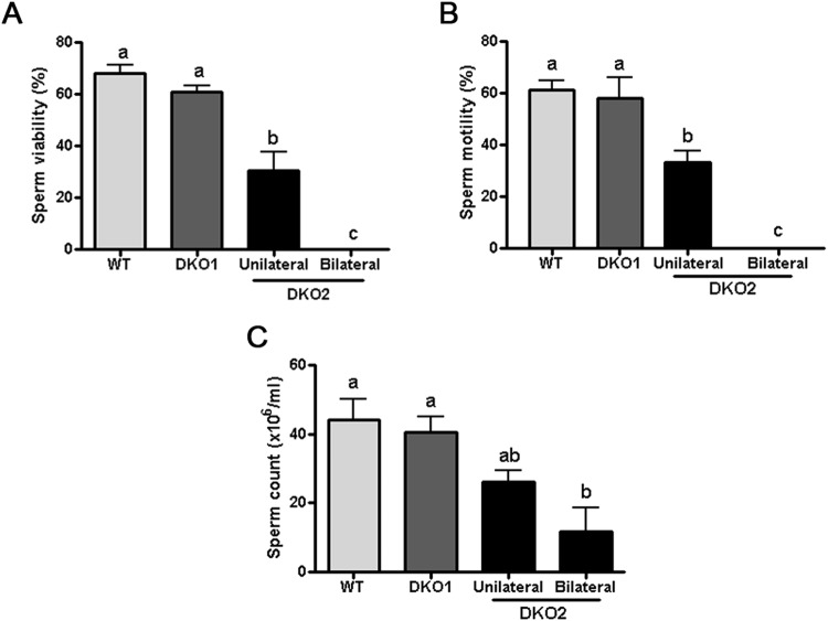
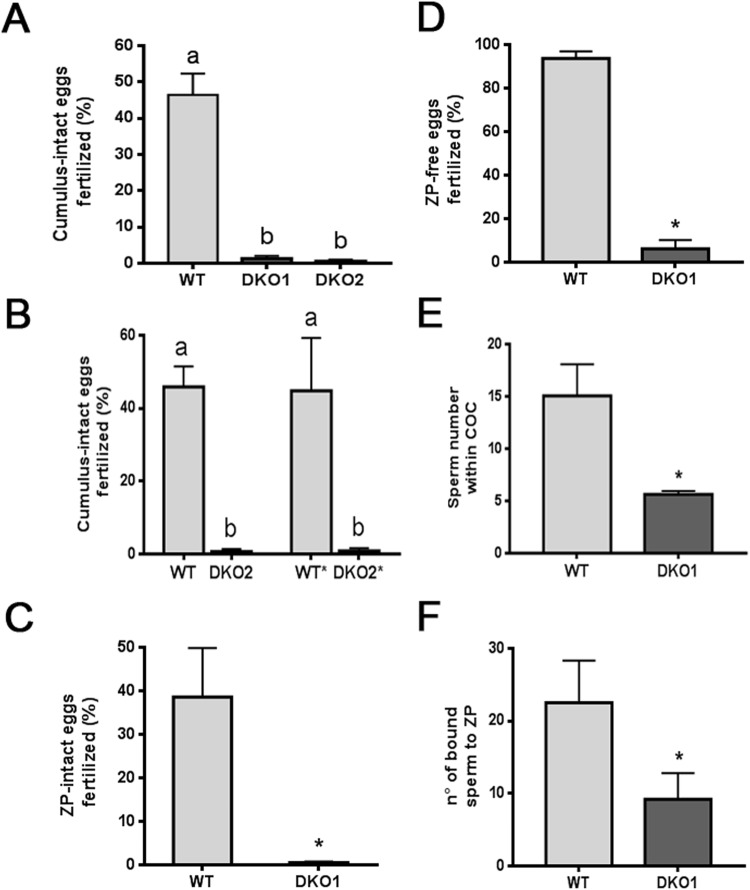
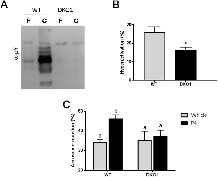
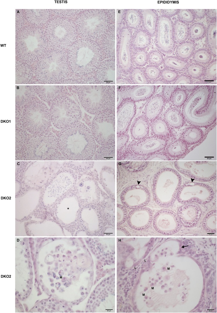
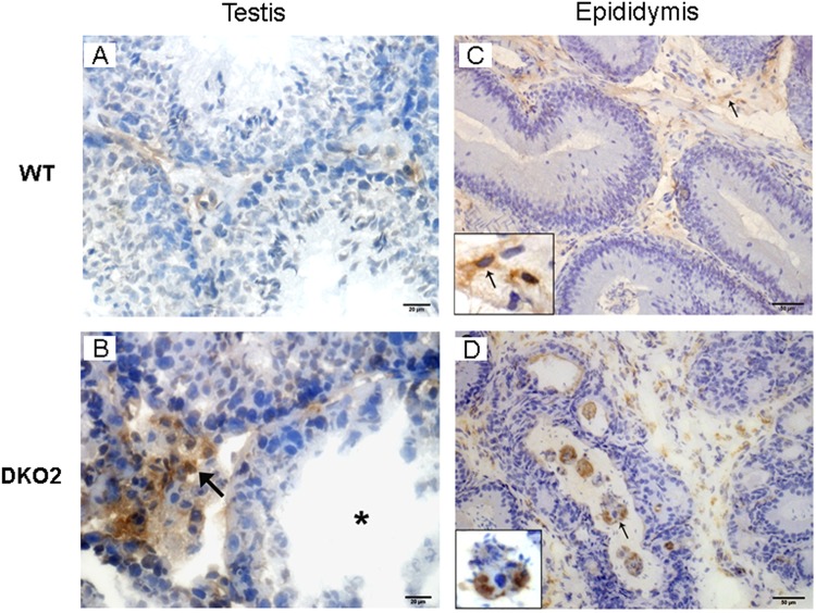
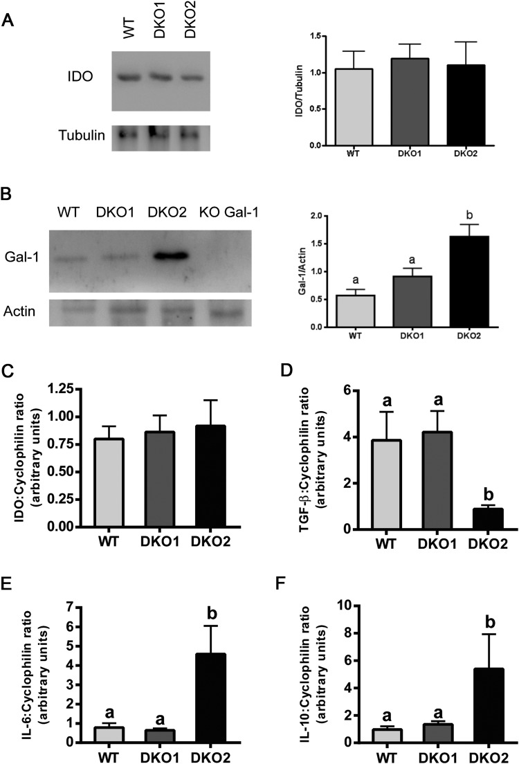
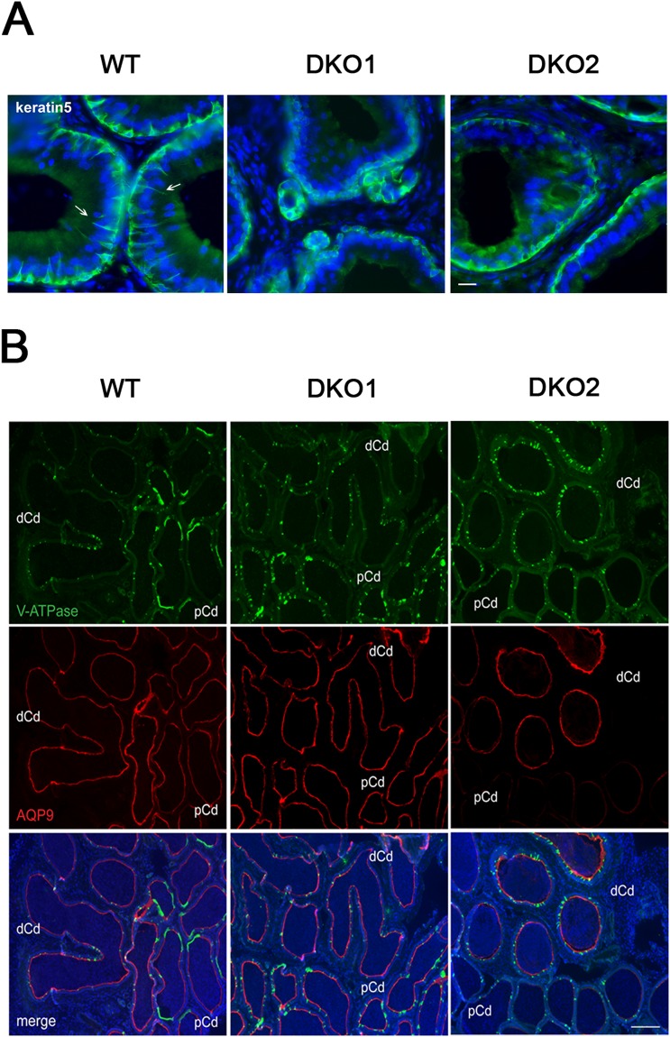
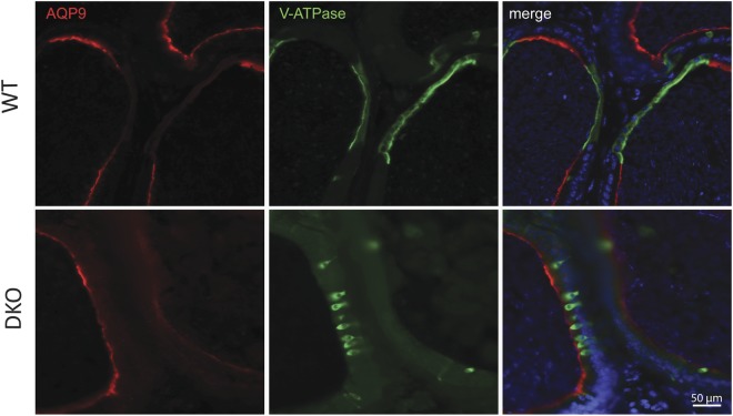
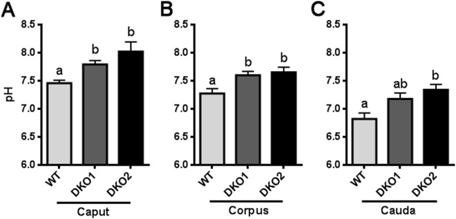
Epididymal Cysteine Rich Secretory Proteins 1 and 4 (CRISP1 and CRISP4) associate with sperm during maturation and play different roles in fertilization. However, males lacking each of these molecules individually are fertile, suggesting compensatory mechanisms between these homologous proteins. Based on this, in the present work, we generated double CRISP1/CRISP4 knockout (DKO) mice and examined their reproductive phenotype. Our data showed that the simultaneous lack of the two epididymal proteins results in clear fertility defects. Interestingly, whereas most of the animals exhibited specific sperm fertilizing ability defects supportive of the role of CRISP proteins in fertilization, one third of the males showed an unexpected epididymo-orchitis phenotype with altered levels of inflammatory molecules and non-viable sperm in the epididymis. Further analysis showed that DKO mice exhibited an immature epididymal epithelium and abnormal luminal pH, supporting these defects as likely responsible for the different phenotypes observed. These observations reveal that CRISP proteins are relevant for epididymal epithelium differentiation and male fertility, contributing to a better understanding of the fine-tuning mechanisms underlying sperm maturation and immunotolerance in the epididymis with clear implications for human epididymal physiology and pathology.


Conflict of interest statement

The authors declare no competing interests.

Figures

Figure 1
Figure 1
Effect of the lack of CRISP1 and CRISP4 on male fertility and in vivo sperm fertilizing ability: (A) WT or DKO adult males were bred with control females for 4 days and the number of pups was analyzed. Data are mean ± SEM; the number of males analyzed (n) is specified in brackets. Different letters indicate significant differences between groups (a vs b, p < 0.01). (B) WT and DKO males were mated with natural estrous females and the percentage of fertilized eggs recovered from the ampulla was evaluated the following day. Data are mean ± SEM; the number of males analyzed (n) is specified in brackets. *p < 0.05. (C) Correlation between the number of pups and the number of fertilized eggs recovered from the ampulla corresponding to females mated by the same males; n = 9, *p < 0.01.
Figure 2
Figure 2
Testicular and epididymal phenotype in DKO mice: (A) representative pictures of DKO and WT epididymides (left) and testes (right). Testicular (B) and epididymal (C) weight relative to total body weight from WT, Group 1 (DKO1) and Group 2 (DKO2) mice. Data are mean ± SEM, n = 21. Caput (D), corpus (E) and cauda (F) weight relative to total body weight from WT, DKO1 and DKO2. Data are mean ± SEM, n = 9. In all cases, different letters indicate significant between groups (a vs b, p < 0.01).
Figure 3
Figure 3
Fertility and in vivo sperm fertilizing ability in each DKO group: (A) WT males as well as DKO mice from Group 1 (DKO1) and from Group 2 (DKO2) were bred with control females for 4 days and the number of pups was analyzed. (B) The same males were mated with natural estrus females and the percentage of fertilized eggs recovered from the ampulla was evaluated the following day. Data are mean ± SEM; the number of males analyzed (n) is specified in brackets. Means not sharing a same letter are significantly different. a vs b, p < 0.01; b vs c and a vs c p < 0.0001.
Figure 4
Figure 4
Effect of the lack of CRISP1 and CRISP4 on fresh sperm parameters. (A) sperm viability was determined by analyzing eosin-labelled cauda epididymal sperm under a light microscope (x400). (B) progressive motility was analyzed in fresh cauda epididymal sperm under light microscope (x400). (C) sperm count was determined using a Neubauer chamber over media containing cauda epididymal sperm obtained by swim out. Data are mean ± SEM, n = 10. Means not sharing a same letter are significantly different. a vs b, p < 0.05; b vs c and a vs c p < 0.0001).
Figure 5
Figure 5
Effect of the lack of CRISP1 and CRISP4 on in vitro sperm fertilizing ability: (A–D) Capacitated sperm from WT or DKO Group 1 (DKO1) or Group 2 (DKO2) males were co-incubated with COC for 3 h (A,B), ZP-intact eggs for 3 h (C) or ZP-free eggs for 1 h (D). At the end of all incubations, fertilization was evaluated by the presence of decondensing sperm heads within the egg cytoplasm. In (B) WT* corresponds to WT capacitated sperm treated to reach the low viability rates of Group 2 DKO sperm and DKO2* represents capacitated sperm from Group 2 DKO mice subjected to a swim up procedure to select viable sperm. Data are mean ± SEM, n = 4. Different letters indicate significant differences between groups (a vs b, p < 0.001). (E) Hoescht-stained capacitated sperm were co-incubated with COC for 15 min and the number of sperm within the cumulus matrix was determined. Data are mean ± SEM, n = 4, *p < 0.05. (F) Capacitated sperm were co-incubated with ZP-intact eggs for 30 min and the number of sperm bound to the ZP was evaluated. Data are mean ± SEM, n = 4, *p < 0.05.
Figure 6
Figure 6
Effect of the lack of CRISP1 and CRISP4 on sperm capacitation-associated events: WT and DKO sperm from Group1 (DKO1) were incubated under capacitating conditions for 90 min and different functional parameters evaluated. (A) Protein tyrosine phosphorylation analyzed by western blotting using an anti-phosphotyrosine antibody (α-pY). Fresh (F) and capacitated (C) sperm were analyzed. (A) representative blot is shown, n = 4. (B) Percentage of hyperactivation evaluated by CASA. Data are mean ± SEM, n = 6, *p < 0.05. (C) Percentage of acrosome reaction determined by Coomassie Brilliant Blue staining in sperm exposed to progesterone (P4) or dimethyl sulfoxide alone (vehicle) during the last 15 min of incubation. Data are mean ± SEM, n = 10. Different letters indicate significant differences between groups (a vs b, p < 0.01).
Figure 7
Figure 7
Histopathology of DKO testes and epididymides: Representative microphotographs of paraffin testis (A–D) and distal caput epididymis (E–H) sections from WT, Group 1 (DKO1) and Group 2 (DKO2) DKO mice stained with hematoxylin-eosin. Normal histology is seen in WT mice (A,E) and DKO1 mice (B,F). In contrast, testis and epididymis sections from DKO2 mice exhibited lymphomononuclear cell infiltrate in lumen and insterstitium (C,D,H) (L: lymphocytes and M: macrophages). Foci of seminiferous tubules severely damaged showing sloughing of germ cells, aspermatogenesis (asterisk) and atrophy (C). In epididymis of DKO2 mice, damage is represented by epithelial cytoplasmic vacuolization (G, arrowheads) and thinning of the epididymal epithelium in a tubule also showing evaginations (H, arrow) In all cases, DKO2 correspond to the enlarged organs from either unilateral- or bilaterally affected mice.
Figure 8
Figure 8
Analysis of macrophages in testis and epididymis: Immunoperoxidase technique using F4/80 antibody to detect macrophages was applied to cryostat testis (A,B) and epididymis (C,D) sections from WT mice, or DKO mice from Group 1 (DKO1) or Group 2 (DKO2). Similar results were observed between WT and DKO1 mice. Representative images of WT mice (A,C) show scarce number of F4/80+ cells scattered in the interstitium. In contrast, DKO2 mice (B,D) show numerous F4/80+ cells in the testis interstitium (B, arrow) close to a damaged seminiferous tubule (B, asterisk) and in the lumen of epididymal tubules with spermatozoa heads within the macrophage cytoplasm (D, arrow). DKO2 correspond to the enlarged organs from either unilateral- or bilaterally affected mice.
Figure 9
Figure 9
Analysis of molecules associated with immune-inflammatory response in DKO epididymides: Expression of immunomodulator proteins IDO (A) and Gal-1 (B) were analyzed by western blotting in epididymides from WT, Group 1 (DKO1) and Group 2 (DKO2) males and relativized to housekeeping proteins. Expression of genes corresponding to different pro- and anti-inflammatory molecules IDO (C), TGF-β (D), IL-6 (E) and IL-10 (F) were evaluated by RT-PCR in epididymides from WT, Group 1 (DKO1) and Group 2 (DKO2) males and relativized to cyclophilin expression. Data are mean ± SEM, n = 6. Different letters indicate significant differences between groups (a vs b, p < 0.05). In all cases, DKO2 values correspond to the inflamed organs from unilateral- or bilaterally affected mice. Full-length blots/gels are presented in Supplementary File.
Figure 10
Figure 10
Effect of the lack of CRISP1 and CRISP4 on epididymal epithelium cells: (A) Immunolabeling of keratin 5 (green), a marker of basal cells, in the initial segment of WT, and DKO from Group 1 (DKO1) and Group 2 (DKO2) epididymides. Basal cells in WT tissues present luminal reaching axiopodia (arrows) not observed in DKO groups. Nuclei are labeled with DAPI (blue). Scales correspond to 10 µm. (B) Double immunolabeling of V-ATPase B1 subunit, a marker of clear cells (green) and AQP9, a marker of principal cells (red) in the cauda epididymis of WT and DKO males. AQP9 staining was restricted to the apical membrane of principal cells (merge image). Clear adjacent cells showed positive staining to V-ATPase in the apical membrane only in WT (merge image). Nuclei are labeled with DAPI (blue). pCd: proximal cauda; dCd: distal cauda. DKO2 correspond to the enlarged organs from unilateral- or bilaterally affected mice. Scales correspond to 100 µm. Representative images are shown.
Figure 11
Figure 11
Effect of the lack of CRISP1 and CRISP4 on cauda epididymal epithelium. Double immunolabeling of AQP9, marker of principal cells (red) and V-ATPase B1 subunit, marker of clear cells (green) in the proximal cauda epididymis of WT and DKO males. Note the more packed together and immature narrow phenotype of clear cells in DKO (similar to pre-pubertal mice) and the absence of the characteristic rows of clear cells observed in WT. Nuclei are labeled with DAPI (blue). Representative images are shown. DKO2 correspond to the enlarged organs from either unilateral- or bilaterally affected mice.
Figure 12
Figure 12
Effect of the lack of CRISP1 and CRISP4 on epididymal luminal pH: Epididymal pH was measured in the luminal content of caput (A), corpus (B) and cauda (C) epididymal regions of WT mice and DKO males from Group 1 (DKO1) or from Group 2 (DKO2). DKO2 values correspond to the inflamed organs from unilateral- or bilaterally affected mice. Data are mean ± SEM, n = 6. Means not sharing a same letter are significantly different. a vs b, p < 0.01; b vs c and a vs c p < 0.0001.

References

    1. Bedford JM. Effects of duct ligation on the fertilzing ability of spermatozoa from different regions of the rabbit epididymis. J. Exp. Zool. 1967;166:271–281. doi: 10.1002/jez.1401660210. - DOI - PubMed
    1. Orgebin-Crist MC. Studies on the function of the epididymis. Biol. Reprod. 1969;1(Suppl 1):155–75. doi: 10.1095/biolreprod1.Supplement_1.155. - DOI - PubMed
    1. Yanagimachi, R. In The Physiologyof Reproduction (eds Knobil, E. & Neill, J. D.) 189–317 (1994).
    1. Cornwall GA. New insights into epididymal biology and function. Hum. Reprod. Update. 2008;15:213–227. doi: 10.1093/humupd/dmn055. - DOI - PMC - PubMed
    1. Sullivan R, Mieusset R. The human epididymis: its function in sperm maturation. Hum. Reprod. Update. 2016;22:574–587. doi: 10.1093/humupd/dmw015. - DOI - PubMed

Publication types

Substances