. 2019 Jan;20(1):97-108.
doi: 10.1038/s41590-018-0260-6. Epub 2018 Dec 3.

The establishment of resident memory B cells in the lung requires local antigen encounter

Affiliations

The establishment of resident memory B cells in the lung requires local antigen encounter

S Rameeza Allie et al. Nat Immunol. 2019 Jan.

Abstract

Memory B cells are found in lymphoid and non-lymphoid tissues, suggesting that some may be tissue-resident cells. Here we show that pulmonary influenza infection elicited lung-resident memory B cells (BRM cells) that were phenotypically and functionally distinct from their systemic counterparts. BRM cells were established in the lung early after infection, in part because their placement required local antigen encounter. Lung BRM cells, but not systemic memory B cells, contributed to early plasmablast responses following challenge infection. Following secondary infection, antigen-specific BRM cells differentiated in situ, whereas antigen-non-specific BRM cells were maintained as memory cells. These data demonstrate that BRM cells are an important component of immunity to respiratory viruses such as influenza virus and suggest that vaccines designed to elicit BRM cells must deliver antigen to the lungs.


Conflict of interest statement

Competing interests statement.

The authors do not have any competing financial or non-financial interests as defined by Nature Research.

Figures

Fig. 1.
Fig. 1.. Identification of influenza–specific B cells.
a. Schematic of recombinant NP and HA proteins. b. Coomassie–stained SDS–PAGE gel showing recombinant HA(PR8) (lane 2), HA(X31) (lane 3) and NP (lane 4). Image is representative of 12 independent preparations of recombinant protein. c–f. Cells from the mLNs of day 15 influenza–infected mice were first gated on live, singlet, CD19+ lymphocytes (Supplementary Fig. 1a), and then on PNA+CD95+ GC B cells (c–d), or first gated on live, singlet, lymphocytes (Supplementary Fig. 1a) and then on CD138+ plasmablasts (e), or first gated on live, singlet, CD19+IgD lymphocytes (Supplementary Fig. 1b) and then on PNAloCD38+ memory B cells (f). Data are representative of 4 independent experiments with 5 mice. Cells from the mLNs of naïve mice (g), day 23 PR8–infected mice (h), and week 9 S. Mansoni–infected mice (i) were gated on live, singlet, CD19+CD38hi lymphocytes (Supplementary Fig. 1c) and analyzed for NP–specific and HA–specific isotype-switched (ISW) memory B cells. Data are representative of 4 experiments with 5 mice.
Fig. 2.
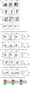
Fig. 2.. Phenotype of memory B cells in the lung and lymphoid organs.
PR8–infected mice were infused with anti–B220 5 minutes prior to euthanasia and cells from the mLN, lung and spleen were gated on live, singlet, lymphocytes (Supplementary Fig. 1a) and then on B220 non–circulating B cells (a) CD38+ memory B cells (b), IgDIgM ISW cells (c) and NP–specific cells (d). Within the population of NP–specific memory cells described in panel d, we determined the frequencies of CD73+PD–L2+ memory B cells (e), CD80PD–L2+ memory B cells (f), CD69+CD103 memory B cells (g), CD62L+CD69+ memory B cells (h), and CXCR3+ memory B cells (i). Data are representative of 3 independent experiments with 5 mice (a–d), 2 experiments with 5 mice (g–h) or 5 experiments with 5 mice. (i). Graphs show individual data points as well as mean ± SD. Data were analyzed by one–way ANOVA with Tukey’s post–test for multiple comparison, ****p=0.0001 ####p=0.0001 (e), ****p=0.0001 ****p=0.0001 (f), ***p=0.0002 *p=0.0105 (g), *p=0.0471 **p=0.0042 (h), *p=0.0206 ***p=0.0008 (i). p<0.05 is considered significant. Cells from the mLN, lung and spleens of day 44 PR8–infected mice were gated on live, singlet, lymphocytes (Supplementary Fig. 1a), and then gated on NP–specific CD19+CD38+IgD memory B cells (Supplementary Fig. 3c–e) and the frequency of IgA, IgG1, IgG2b, IgG2c, IgG3 and IgM–expressing cells was determined (j). Data are representative of 3 independent experiments with 5 mice.
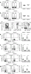
Fig. 3.
Fig. 3.. Identification of influenza–specific, non–circulating BRM cells in the lung.
Mice were infected on day 0, surgically paired with partner mice on day 44 and analyzed on day 59 (a–i). Cells from the mLN (a) or spleen (b) of each partner mouse were gated on live, singlet, lymphocyte CD19+CD38+IgD+IgMlo naïve B cells (Supplementary Fig. 1d) and the frequency of CD45.2+ cells was determined in each partner. Data are combined from 4 independent experiments with 3 mice each. Graph shows individual points as well as mean ± SD. Cells from the lung were gated on live, singlet, lymphocytes (Supplementary Fig. 1a) and subsequently gated on CD19+B220 (non–circulating), CD38+IgDIgM+ (IgM) or CD38+IgDIgM (ISW) memory B cells (c). The frequencies and numbers of NP–specific IgM (d) and ISW (e) memory B cells from host and partner mice were determined in the lungs of naïve mice paired with PR8–infected mice. The frequencies and numbers of NP–specific IgM (f) and ISW (g) memory B cells from host and partner mice were determined in the lungs of LPS–treated mice paired with PR8–infected mice. The frequencies and numbers of NP–specific IgM (h) and ISW (i) memory B cells from host and partner mice were determined in the lungs of PR8–infected mice paired with PR8–infected mice. Data are representative of 3 independent experiments, each with 3 pairs of mice (d–e), 2 experiments with 3 pairs of mice (f–g), or 3 experiments combined, totaling 11 pairs of mice (h–i). Graphs show individual data as well as mean ± SD. Significance was determined using one–way ANOVA (paired) followed by the Bonferroni–Sidak method for multiple comparison, *p=0.0428 (d), ***p=0.0001 (e–f), ****p=0.0001, **p=0.0040 (h), ****p=0.0001 **p=0.0022 (i). p<0.05 is considered significant.
Fig. 4.
Fig. 4.. BRM cells in the lung are established early after infection.
Mice were infected on day 0, surgically paired with partner mice on day 15 and analyzed on day 30 (a–g). Cells from the mLN (a) or spleen (b) were gated on live, singlet, lymphocyte CD19+IgD+IgMlo naïve B cells (Supplementary Fig. 1d) and the frequency of CD45.2+ cells was determined in each partner. These data are combined from 2 independent experiments totaling 8 pairs of mice. Graphs show individual data points as well as mean ± SD. Cells from the lung were gated on live, singlets (Supplementary Fig. 1a) and subsequently gated on live, CD19+B220 (non–circulating), CD38+IgDIgM+ (IgM) or CD38+IgDIgM (ISW) memory B cells (c). The frequencies of NP–specific IgM (d) and ISW (e) memory B cells from the lungs of host and partner mice. The frequencies of HA(PR8)–specific IgM (f) and ISW (g) memory B cells from the lungs of host and partner mice. Data in c–g are representative of 3 independent experiments, each with 4 pairs of mice. Graphs show individual data as well as mean ± SD. Significance was determined using one–way ANOVA (paired) followed by the Bonferroni–Sidak method for multiple comparison, *p=0.0457 ***p=0.0002 (e), **p=0.0047 *p=0.0213 (f), *p=0.0285 ***p=0.0002 (g), **p=0.0060 ##p=0.0080 (h). p<0.05 is considered significant.
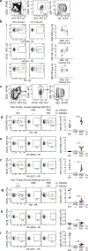
Fig. 5.
Fig. 5.. BRM cells in the lung are generated from early CD40–dependent precursors.
Cells from the mLN of PR8–infected mice were gated on live, singlet, lymphocyte, CD19+PNA+CD95+ GC B cells (Supplementary Fig. 1f) and NP–specific as well as HA–specific cells were enumerated (a). Cells from the blood (b) and lungs (c) of PR8–infected mice were gated on live, singlet, lymphocyte, CD19+CD38+IgMIgD ISW memory B cells (Supplementary Figure 1c), and the frequency (b) and number (c) of NP–specific as well as HA–specific cells was determined. Data are representative of 3 independent experiments with 5 mice at each timepoint. The data points represent mean ± SD. Mice were infected with PR8, administered anti–CD40L (MR1) or isotype control antibody (CT) every other day for 10 days starting on day 5 (d, h), day 10 (e, i), day 20 (f, j) or day 30 (g, k). Cells from the mLN and lung were gated on live, singlet, lymphocyte, CD19+CD138lo cells (Supplementary Fig. 1g) and subsequently gated on GL7+CD38lo GC B cells (d–g). Cells from the lung were gated on live, singlet, lymphocyte, CD19+CD138loCD38+IgM+IgD IgM BRM cells or CD19+CD138loCD38+IgMIgD ISW BRM cells (h–k)(gating in Supplementary Fig. 1g). Data in d–k are representative of 3 independent experiments with 5 mice/group/timepoint. Graphs show mean ± SD as well as individual data points. Significance was determined using a Mann–Whitney U test, **p=0.0079 ##p=0.0014 (d), **p=0.0079 *p=0.0462 (e), **p=0.0079, ##p=0.0024 (f) *p=0.0159 #p=0.0180 (g) or unpaired, two–tailed t–test **p=0.0018, ***p=0.0003 (h), **p=0.0011 (i). p<0.05 is considered significant.
Fig. 6.
Fig. 6.. Establishment of BRM cells in the lung requires local antigen encounter.
Naïve mice were surgically joined and, on day 15, one was infected with PR8 and the other infected with X31 and BRM cells in the lung analyzed on day 25 (a–b). Cells from the lung were gated on live, singlet, lymphocytes (Supplementary Fig 1a), and subsequently gated on B220CD19+CD38hiIgMIgD ISW BRM cells (a). The frequency and number of HA(PR8)–specific (top row), HA(X31)–specific (middle row) and NP–specific (bottom row) BRM cells were determined in each partner (b). Data are representative of 3 independent experiments with 5 pairs of mice. Significance was determined using one–tailed, paired t test, ****p=0.0001, *p=0.0477. Mice were peritoneally–infected with PR8 on day 0, intranasally challenged with X31 on day 30 and analyzed on days 40 (d–f) and day 75 (g–i). Cells from the lung were gated on live, singlet, lymphocytes (Supplementary Fig. 1a) and subsequently gated on CD19+B220CD38+IgDIgM ISW memory B cells (c). NP–specific (d,g), HA(PR8)–specific (e,h) and HA(X31)–specific (f,i) ISW BRM cells were enumerated on day 40 (d–f) and day 75 (g–i). These data are representative of 2 independent experiments with 5 mice/timepoint. Data were analyzed with an unpaired t test, ****p=0.0001 (f), ***p=0.0009 (g), **p=0.0013 (h), **p=0.0047 (i), **p=0.0011 (j), ***p=0.0004 (k). p<0.05 is considered significant.
Fig. 7.
Fig. 7.. BRM cells are associated with protection from secondary infection.
Mice were infected in the peritoneal cavity or in the lung and analyzed on day 30 (a–d). Cells were gated on live, singlet, lymphocyte, CD19+B220CD138GL7+CD38lo GC B cells (Supplemental Fig. 1h) and NP–specific cells were enumerated in the spleen (a). Cells were gated on live, singlet, lymphocyte CD19+B220CD38+IgDIgM isotype-switched memory B cells (Supplemental Fig. 1e) and NP–specific cells were enumerated in the spleen (b) and lung (c). Data are representative of 3 independent experiments with 3 mice in the naïve group, 5 mice in the i.p. infected group and 4 mice in the i.n. infected group. Graphs show mean ± SD as well as individual data points. Significance was determined with a one–way ANOVA with Tukey’s post–test for multiple comparisons, **p=0.0038 (a), **p=0.0021 (b), ****p=0.0001 (c). Weight loss after challenge infection (d). Viral titers on day 5 after challenge infection (e). Data in d, e are representative of 2 experiments with 5 mice/group. Graphs show mean ± SD (d) or individual data points as well as the geometric mean ± SD (e). Data were analyzed using the Mann–Whitney U test, **p=0.0079. p<0.05 is considered significant.
Fig. 8.
Fig. 8.. BRM cells are required for rapid secondary ASCs in the lung.
a. NP–specific ELISPOTs in the lung 4 days after challenge infection. Graph shows data points as well as mean ± SD. Data are representative of 5 independent experiments with 4 mice/group. Data were analyzed with one–way ANOVA and Tukey’s test for multiple comparisons, **p=0.0037, ##p=0.002, $ $p=0.003. b–c. BLIMP–1–reporter mice were infected with PR8 on day 0, challenged with X31 on day 30 (b) or 60 (c) and ASCs were enumerated 3 days later. Cells in the lung were gated on live, singlet, lymphocytes (Supplementary Fig. 1a) and subsequently gated on NP–specific YFP+ cells. Data are representative of 3 independent experiments (b) or 2 experiments (c) with 3–5 mice/group. Graphs show individual data points as well as mean ± SD. Data are analyzed by 1–way ANOVA with Bonnferoni–Sidak test for multiple comparisons, ***p=0.0003, ****p=0.0001, ####p=0.0001 (b) or Tukey’s test for multiple comparisons, ****p=0.0001 ####p=0.0001 $ $ $ $p=0.0001 (c). d. Mice were infected with PR8 on day 0, treated with FTY720, challenged with X31 on day 30 and ELISPOTs in the lung were analyzed 4 days later. Data are representative of 2 independent experiments with 6 mice/group (control) or 4 mice/group (FTY720). Graph shows data points as well as mean ± SD. Data were analyzed with a Mann–Whitney U test. e–f. Mice were infected with PR8 on day 0, challenged with X31 on day 30 and analyzed on day 33. Cells from the lung were gated on live, singlet, lymphocyte, CD19+B220CD38+IgDIgM ISW memory B cells (Supplementary Fig. 1e) and NP–specific (e) HA(PR8)–specific (f) memory B cells were enumerated. Graphs show individual data points as well as mean ± SD. Data are representative of 3 independent experiments with 3 mice/group. Data were analyzed with Student’s t test, *p=0.0488. p<0.05 is considered significant.

Comment in

  • Recent Advances in Lung Immunobiology.
    Ramonell RP, Prasla Z, Terry CR, Schulman DA, Lee FE. Ramonell RP, et al. Am J Respir Cell Mol Biol. 2019 Dec;61(6):786-788. doi: 10.1165/rcmb.2019-0183RO. Am J Respir Cell Mol Biol. 2019. PMID: 31291124 Free PMC article. No abstract available.

References

    1. Halliley JL et al. Long-Lived Plasma Cells Are Contained within the CD19-CD38hiCD138+ Subset in Human Bone Marrow. Immunity 43, 132–145 (2015). - PMC - PubMed
    1. Dorner T & Radbruch A Antibodies and B cell memory in viral immunity. Immunity 27, 384–392 (2007). - PubMed
    1. Phan TG & Tangye SG Memory B cells: total recall. Curr. Opin. Immunol 45, 132–140 (2017). - PubMed
    1. Bannard O & Cyster JG Germinal centers: programmed for affinity maturation and antibody diversification. Curr. Opin. Immunol 45, 21–30 (2017). - PubMed
    1. Shlomchik MJ & Weisel F Germinal center selection and the development of memory B and plasma cells. Immunol. Rev 247, 52–63 (2012). - PubMed

Publication types

MeSH terms