Enhancement of wound healing by single-wall/multi-wall carbon nanotubes complexed with chitosan
- PMID: 30510412
- PMCID: PMC6231507
- DOI: 10.2147/IJN.S183342
Enhancement of wound healing by single-wall/multi-wall carbon nanotubes complexed with chitosan
Abstract
Background: Impaired wound healing is commonly associated with many health problems, including diabetes, bedsores and extensive burns. In such cases, healing often takes a long time, which subjects patients to various complications. This study aims to investigate whether single-wall or multi-wall carbon nanotubes complexed with chitosan hydrogel can improve wound healing.
Materials and methods: Initially, the effects of the complexes on the viability and functionality of fibroblasts were investigated in engineered connective tissues. Then, their activity on wound healing was investigated in a mouse model with induced full-thickness wounds, in which the wounds were treated daily with these complexes. Finally, the effect of the complexes on collagen deposition by fibroblasts was investigated in vitro.
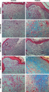
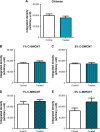
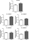
Results: The engineered connective tissue studies showed that fibroblasts were viable in the presence of the complexes and were still able to effectively organize and contract the extracellular matrix. In vivo data showed that both types of complexes improved the re-epithelialization of the healing wounds; however, they also increased the percentage of wounds with higher fibrosis. In particular, the chitosan-multi-wall carbon nanotube complex significantly enhanced the extensiveness of this fibrosis, which is in line with in vitro data showing a concentration-dependent enhancement of collage deposition by these complexes. These observations were associated with an increase in inflammatory signs in the wound bed.
Conclusion: Single-wall and multi-wall carbon nanotubes complexed with chitosan improved the re-epithelialization of wounds, but an increase in fibrosis was detected.
Keywords: carbon nanotubes; chitosan; fibrosis; inflammation; re-epithelialization; wound healing.
Conflict of interest statement
Disclosure The authors report no conflicts of interest in this work.
Figures


Similar articles
-
SIKVAV-Modified Chitosan Hydrogel as a Skin Substitutes for Wound Closure in Mice.Molecules. 2018 Oct 11;23(10):2611. doi: 10.3390/molecules23102611. Molecules. 2018. PMID: 30314388 Free PMC article.
-
Dressing with epigallocatechin gallate nanoparticles for wound regeneration.Wound Repair Regen. 2016 Mar;24(2):287-301. doi: 10.1111/wrr.12372. Epub 2015 Dec 10. Wound Repair Regen. 2016. PMID: 26472668
-
Evaluation of wound-healing efficiency of a functional Chitosan/Aloe vera hydrogel on the improvement of re-epithelialization in full thickness wound model of rat.J Tissue Viability. 2022 Nov;31(4):649-656. doi: 10.1016/j.jtv.2022.07.009. Epub 2022 Jul 31. J Tissue Viability. 2022. PMID: 35965210
-
Assessing Animal Models to Study Impaired and Chronic Wounds.Int J Mol Sci. 2024 Mar 29;25(7):3837. doi: 10.3390/ijms25073837. Int J Mol Sci. 2024. PMID: 38612647 Free PMC article. Review.
-
Fibroblasts-Warriors at the Intersection of Wound Healing and Disrepair.Biomolecules. 2023 Jun 6;13(6):945. doi: 10.3390/biom13060945. Biomolecules. 2023. PMID: 37371525 Free PMC article. Review.
Cited by
-
Cell Therapy and Investigation of the Angiogenesis of Fibroblasts with Collagen Hydrogel on the Healing of Diabetic Wounds.Turk J Pharm Sci. 2023 Nov 7;20(5):302-309. doi: 10.4274/tjps.galenos.2022.62679. Turk J Pharm Sci. 2023. PMID: 37933815 Free PMC article.
-
Nanomedicine and Its Role in Surgical Wound Infections: A Practical Approach.Bioengineering (Basel). 2025 Jan 31;12(2):137. doi: 10.3390/bioengineering12020137. Bioengineering (Basel). 2025. PMID: 40001657 Free PMC article. Review.
-
Multifunctional Carbon-Based Nanocomposite Hydrogels for Wound Healing and Health Management.Gels. 2025 May 6;11(5):345. doi: 10.3390/gels11050345. Gels. 2025. PMID: 40422365 Free PMC article. Review.
-
State of the Art in the Antibacterial and Antiviral Applications of Carbon-Based Polymeric Nanocomposites.Int J Mol Sci. 2021 Sep 29;22(19):10511. doi: 10.3390/ijms221910511. Int J Mol Sci. 2021. PMID: 34638851 Free PMC article. Review.
-
A critical overview of challenging roles of medicinal plants in improvement of wound healing technology.Daru. 2024 Jun;32(1):379-419. doi: 10.1007/s40199-023-00502-x. Epub 2024 Jan 15. Daru. 2024. PMID: 38225520 Free PMC article.
References
-
- Neville RF, Kayssi A, Buescher T, Stempel MS. The diabetic foot. Curr Probl Surg. 2016;53(9):408–437. - PubMed
-
- O’Meara S, Cullum N, Majid M, Sheldon T. Systematic reviews of wound care management: (3) antimicrobial agents for chronic wounds; (4) diabetic foot ulceration. Health Technol Assess. 2000;4(21):1–237. - PubMed
-
- Değim Z, Celebi N, Sayan H, Babül A, Erdoğan D, Take G. An investigation on skin wound healing in mice with a taurine-chitosan gel formulation. Amino Acids. 2002;22(2):187–198. - PubMed
-
- Kong M, Chen XG, Xing K, Park HJ. Antimicrobial properties of chitosan and mode of action: a state of the art review. Int J Food Microbiol. 2010;144(1):51–63. - PubMed
MeSH terms
Substances
LinkOut - more resources
Full Text Sources

