. 2018 Oct 29:2018:7453161.
doi: 10.1155/2018/7453161. eCollection 2018.

Angiotensin II Attenuates the Bioactivities of Human Endothelial Progenitor Cells via Downregulation of β 2-Adrenergic Receptor

Affiliations

Angiotensin II Attenuates the Bioactivities of Human Endothelial Progenitor Cells via Downregulation of β 2-Adrenergic Receptor

Seon Jin Lee et al. Stem Cells Int. .

Abstract

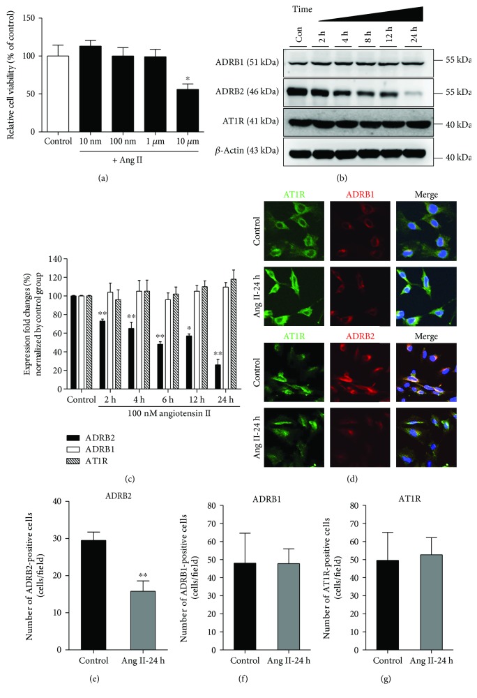
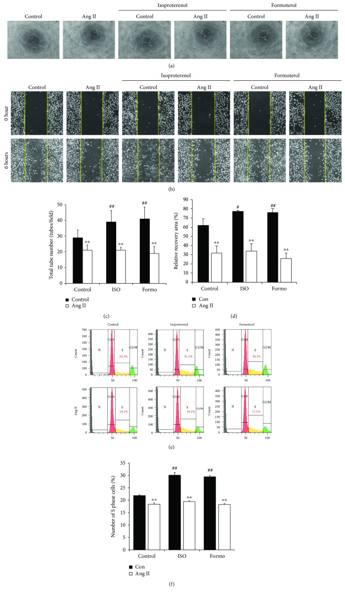
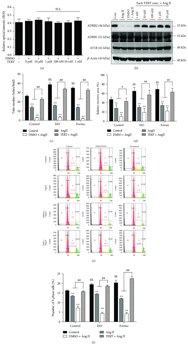
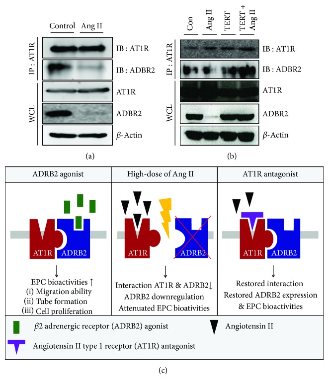
Cross talks between the renin-angiotensin system (RAS), sympathetic nervous system, and vascular homeostasis are tightly coordinated in hypertension. Angiotensin II (Ang II), a key factor in RAS, when abnormally activated, affects the number and bioactivity of circulating human endothelial progenitor cells (hEPCs) in hypertensive patients. In this study, we investigated how the augmentation of Ang II regulates adrenergic receptor-mediated signaling and angiogenic bioactivities of hEPCs. Interestingly, the short-term treatment of hEPCs with Ang II drastically attenuated the expression of beta-2 adrenergic receptor (ADRB2), but did not alter the expression of beta-1 adrenergic receptor (ADRB1) and Ang II type 1 receptor (AT1R). EPC functional assay clearly demonstrated that the treatment with ADRB2 agonists significantly increased EPC bioactivities including cell proliferation, migration, and tube formation abilities. However, EPC bioactivities were decreased dramatically when treated with Ang II. Importantly, the attenuation of EPC bioactivities by Ang II was restored by treatment with an AT1R antagonist (telmisartan; TERT). We found that AT1R binds to ADRB2 in physiological conditions, but this binding is significantly decreased in the presence of Ang II. Furthermore, TERT, an Ang II-AT1R interaction blocker, restored the interaction between AT1R and ADRB2, suggesting that Ang II might induce the dysfunction of EPCs via downregulation of ADRB2, and an AT1R blocker could prevent Ang II-mediated ADRB2 depletion in EPCs. Taken together, our report provides novel insights into potential therapeutic approaches for hypertension-related cardiovascular diseases.


Figures

Figure 1
Figure 1
Effect of Ang II on ADRB2 expression and EPC bioactivities. (a) Ang II-induced cytotoxicity in EPCs was measured using WST-1 assay. EPC viability was reduced after treatment with 10 μM Ang II. P < 0.05 vs. control. (b). ADRB1, ADRB2, and AT1R levels after time-dependent Ang II treatment were analyzed using Western blotting, and β-actin was used as a loading control. (c). Quantitative graph of total protein levels in Ang II-induced EPCs. P < 0.01 and ∗∗ P < 0.001 vs. control. (d) Immunocytochemistry was performed to confirm the expression of ADRB1, ADRB2, and AT1R in the presence of Ang II. Representative cropped images of ADRB1, ADRB2, and AT1R from 20x fluorescent images. (e–g) Quantification of ADRB2-, ADRB1-, and AT1R-positive cells per field. ∗∗ P < 0.01 vs. control.
Figure 2
Figure 2
ADRB2 agonists stimulate EPC bioactivities. (a) Human EPCs were pretreated with Ang II, and the effects of ADRB2 agonists (isoproterenol and formoterol) were analyzed for test tube formation ability. (b) Migration ability was estimated by a scratch wound healing assay. Each group of EPCs, control cells, or Ang II-treated cells was seeded, followed by treatment with media containing ADRB2 agonists. (c) Quantification of total tube number was performed using ImageJ software. All experiments were performed in triplicates. ∗∗ P < 0.01 vs. control, and ## P < 0.01 vs. negative control. (d) Quantification of migrated cells was performed using ImageJ software. Recovery area was analyzed and presented. ∗∗ P < 0.01 vs. control; # P < 0.05 and ## P < 0.01 vs. negative control. (e) Propidium iodide (PI) staining of DNA was detected by flow cytometry of EPCs treated with 100 nM Ang II. (f) Graph of the proportion of cells in the S phase for each group as measured by PI staining. ∗∗ P < 0.01 vs. control, and ## P < 0.01 vs. negative control.
Figure 3
Figure 3
Effects of AT1R blocker, telmisartan (TERT), on EPCs. (a) Cell viability assay upon treatment with TERT, an AT1R blocker, using the WST-1 assay. NS = not significant. (b) Ang II- and TERT-treated EPCs were harvested, and the expression of ADRB1, ADRB2, and AT1R was analyzed using Western blotting. Expression of ADRB2 was decreased following treatment of EPCs with Ang II, whereas protein levels of ADRB2 were restored in AT1R-blocked cells. (c) Human EPCs were pretreated with an AT1R blocker and incubated with Ang II for 24 h. Each group of cells was seeded onto Matrigel GFR with or without ADRB2 agonists. Quantitative graph of the tube formation data. The tube number was measured using ImageJ software. ∗∗ P < 0.01 vs. control, $$ P < 0.01 vs. negative control, and ## P < 0.01 vs. Ang II or DMSO plus Ang II. (d) Migration ability was examined using the scratch wound healing assay. Each group of EPCs was seeded, and migratory capacity was observed for 6 h. Quantitative graph of migration assay results. All experiments were performed in triplicates at least. ∗∗ P < 0.01 vs. control, $$ P < 0.01 vs. negative control, and # P < 0.05 and ## P < 0.01 vs. Ang II or DMSO plus Ang II. (e, f) PI staining of DNA in EPCs detected by flow cytometry. Graph of the percentage of S-phase cells in each group measured by PI staining. ∗∗ P < 0.01 vs. control, $$ P < 0.01 vs. negative control, and ## P < 0.01 vs. Ang II or DMSO plus Ang II.
Figure 4
Figure 4
The interaction between AT1R and ADRB2 is attenuated by Ang II. (a) Association between AT1R and ADRB2 in EPCs was confirmed by immunoprecipitation assay. For endogenous IP, lysate from EPCs was immunoprecipitated using a specific antibody, anti-AT1R, followed by detection with ADRB2 antibody. (b) To confirm the role of Ang II in the interaction between AT1R and ADRB2, EPCs were treated with Ang II or TERT. (c) Proposed working model for the role of ADRB2 in Ang II-induced EPC bioactivities.

References

    1. Manrique C., Lastra G., Gardner M., Sowers J. R. The renin angiotensin aldosterone system in hypertension: roles of insulin resistance and oxidative stress. The Medical Clinics of North America. 2009;93(3):569–582. doi: 10.1016/j.mcna.2009.02.014. - DOI - PMC - PubMed
    1. Kamide K., Rakugi H., Higaki J., et al. The renin-angiotensin and adrenergic nervous system in cardiac hypertrophy in fructose-fed rats. American Journal of Hypertension. 2002;15(1):66–71. doi: 10.1016/S0895-7061(01)02232-4. - DOI - PubMed
    1. Kobori H., Ohashi N., Katsurada A., et al. Urinary angiotensinogen as a potential biomarker of severity of chronic kidney diseases. Journal of the American Society of Hypertension. 2008;2(5):349–354. doi: 10.1016/j.jash.2008.04.008. - DOI - PMC - PubMed
    1. de Cavanagh E. M. V., González S. A., Inserra F., et al. Sympathetic predominance is associated with impaired endothelial progenitor cells and tunneling nanotubes in controlled-hypertensive patients. American Journal of Physiology. Heart and Circulatory Physiology. 2014;307(2):H207–H215. doi: 10.1152/ajpheart.00955.2013. - DOI - PubMed
    1. Nagy J. A., Dvorak A. M., Dvorak H. F. VEGF-A and the induction of pathological angiogenesis. Annual Review of Pathology. 2007;2(1):251–275. doi: 10.1146/annurev.pathol.2.010506.134925. - DOI - PubMed

LinkOut - more resources