Review
. 2018 Oct 28:2018:6578097.
doi: 10.1155/2018/6578097. eCollection 2018.

Traumatic Lumbosacral Dislocation: Current Concepts in Diagnosis and Management

Affiliations
Review

Traumatic Lumbosacral Dislocation: Current Concepts in Diagnosis and Management

Andrew S Moon et al. Adv Orthop. .

Abstract

Traumatic lumbosacral dislocation is a rare, high-energy mechanism injury characterized by displacement of the fifth lumbar vertebra in relation to the sacrum. Due to the violent trauma typically associated with this lesion, there are often severe, coexisting injuries. High-quality radiographic studies, in addition to appropriate utilization of CT scan and MRI, are essential for proper evaluation and diagnosis. Although reports in the literature include nonoperative and operative management, most authors advocate for surgical treatment with open reduction and decompression with instrumentation and fusion. Despite advances in early diagnosis and management, this injury type is associated with significant morbidity and mortality, and long-term patient outcomes remain unclear.


Figures

Figure 1
Figure 1
Classification of fracture-dislocation of the fifth lumbar vertebra according to Aihara et al. [1].
Figure 2
Figure 2
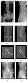
Imaging of a 25-year-old male patient who was involved in an all-terrain vehicle accident. He was ejected from the vehicle and presented with low back pain and intermittent bilateral lower extremity radicular pain with paresthesia. Figures (a) and (b) demonstrate anteroposterior and lateral radiographs, respectively. Coronal CT shows minimal lateralization of L5 over S1 (c). Sagittal view shows anterior dislocation of L5 over S1 with jumped facets (d). Axial (e) image cut through the same level as the sagittal image (f) shows bilateral jumped facets at L5-S1. The patient underwent posterior spinal instrumentation and fusion of the L5 and S1 vertebrae using pedicle screws and rods. Postoperative anteroposterior (g) and lateral (h) images demonstrate a reduced L5-S1 joint. (Courtesy of University of Alabama at Birmingham, Department of Orthopaedic Surgery, Spine Fellowship Program, Birmingham, Alabama, USA).

References

    1. Aihara T., Takahashi K., Yamagata M., Moriya H. Fracture-dislocation of the fifth lumbar vertebra. The Journal of Bone & Joint Surgery (British Volume) 1998;80(5):840–845. doi: 10.1302/0301-620X.80B5.8657. - DOI - PubMed
    1. B. Grivas T. Unilateral Lumbosacral Dislocation: Case Report and a Comprehensive Review. The Open Orthopaedics Journal. 2012;6(1):473–477. doi: 10.2174/1874325001206010473. - DOI - PMC - PubMed
    1. Robbins M., Mallon Z., Roberto R., Patel R., Gupta M., Klineberg E. Traumatic spondylopelvic dissociation: A report of two cases of spondylolisthesis at l5-s1 and review of literature. Global Spine Journal. 2015;5(3):225–230. doi: 10.1055/s-0035-1549435. - DOI - PMC - PubMed
    1. Reinhold M., Knop C., Blauth M. Acute traumatic L5-S1 spondylolisthesis: A case report. Archives of Orthopaedic and Trauma Surgery. 2006;126(9):624–630. doi: 10.1007/s00402-005-0078-1. - DOI - PubMed
    1. Reddy S. J., Al-Holou W. N., Leveque J.-C., La Marca F., Park P. Traumatic lateral spondylolisthesis of the lumbar spine with a unilateral locked facet: Description of an unusual injury, probable mechanism, and management: Report of two cases. Journal of Neurosurgery: Spine. 2008;9(6):576–580. doi: 10.3171/SPI.2008.6.08301. - DOI - PubMed

LinkOut - more resources