

Secondary hyperhomocysteinemia-related occlusive retinal vasculopathy: A case report
- PMID: 30511034
- PMCID: PMC6258140
- DOI: 10.1016/j.ajoc.2018.11.005
Secondary hyperhomocysteinemia-related occlusive retinal vasculopathy: A case report
Abstract
Purpose: To report a case of occlusive retinal vasculopathy, secondary to hyperhomocysteinemia.
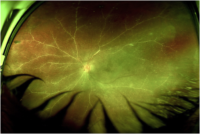
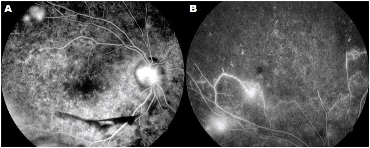
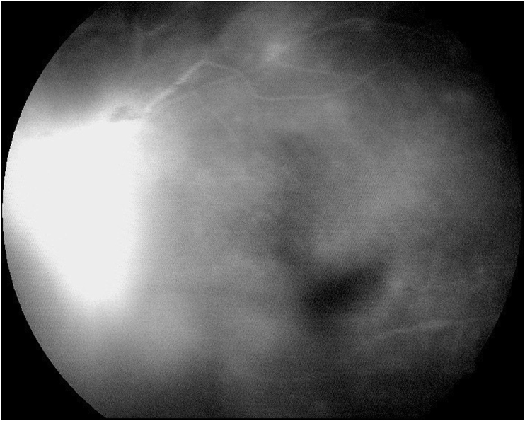
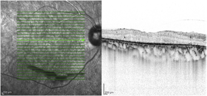
Observations: A 43-year-old male was examined at the retina outpatient clinic due to complaints of bilateral decrease in visual acuity. The patient underwent a comprehensive ophthalmological examination, wide-field fundus photographs and fluorescein angiography, as well as spectral domain optical coherence tomography with enhanced-deep imaging. The patient had a significant medical history of chronic kidney disease and progressive bilateral vision loss over the last two years, which worsened in the left eye during the past 3 months. Fundus examination revealed a vitreous hemorrhage in the left eye and bilateral proliferative retinopathy. Blood glucose and systemic blood pressure were unremarkable. Plasma homocysteine was reported at >500 μmol/L, which is higher than the corrected reference range by age.
Conclusion and importance: Hyperhomocysteinemia is a rare but well-known disease, capable of accelerating atherosclerotic disease and generating a prothrombotic state that can lead to multiple systemic complications. Despite its low incidence, the disease should be part of the differential diagnosis in patients with bilateral proliferative retinopathy, in the absence of diabetes mellitus and systemic hypertension.
Keywords: Atherosclerosis; Chronic kidney disease; Hyperhomocysteinemia; Occlusive retinal vasculopathy; Proliferative retinopathy.
Figures


Similar articles
-
A case of bilateral pachychoroid disease: polypoidal choroidal vasculopathy in one eye and peripheral exudative hemorrhagic chorioretinopathy in contralateral eye.BMC Ophthalmol. 2021 Sep 4;21(1):320. doi: 10.1186/s12886-021-02067-2. BMC Ophthalmol. 2021. PMID: 34481477 Free PMC article.
-
Retinal arterial occlusive vasculitis after multiple intravitreal brolucizumab injections for diabetic macular edema.Am J Ophthalmol Case Rep. 2022 Dec 30;29:101788. doi: 10.1016/j.ajoc.2022.101788. eCollection 2023 Mar. Am J Ophthalmol Case Rep. 2022. PMID: 36632338 Free PMC article.
-
CLINICALLY INVISIBLE RETINAL HEMANGIOBLASTOMAS DETECTED BY SPECTRAL DOMAIN OPTICAL COHERENCE TOMOGRAPHY AND FLUORESCEIN ANGIOGRAPHY IN TWINS.Retin Cases Brief Rep. 2018 Winter;12(1):12-16. doi: 10.1097/ICB.0000000000000382. Retin Cases Brief Rep. 2018. PMID: 27533642
-
Giraffe or leopard spot chorioretinopathy as an outstanding finding: case report and literature review.Int Ophthalmol. 2019 Jun;39(6):1405-1412. doi: 10.1007/s10792-018-0948-5. Epub 2018 Jun 8. Int Ophthalmol. 2019. PMID: 29948498 Review.
-
Proliferative retinopathy during hyperbaric oxygen treatment.Diving Hyperb Med. 2017 Sep;47(3):203. doi: 10.28920/dhm47.3.203. Diving Hyperb Med. 2017. PMID: 28868603 Free PMC article. Review.
References
-
- Koylu M.T., Kucukevcilioglu M., Erdurman F.C. Association of retinal vein occlusion, homocysteine, and the thrombophilic mutations in a Turkish population: a case-control study. Ophthalmic Genet. 2017:1–5. 0(0) - PubMed
-
- Wall R.T., Harlan J.M., Harker L.A., Striker G.E. Homocysteine-induced endothelial cell injury in vitro: a model for the study of vascular injury. Thromb Res. 1980;18(1-2):113–121. - PubMed
-
- Ajith T.A., Ranimenon Homocysteine in ocular diseases. Clin Chim Acta. 2015;450:316–321. - PubMed
-
- Wright A., Martin N., Dodson P. Homocysteine, folates, and the eye. Eye. 2008;22(8):989–993. - PubMed
Publication types
LinkOut - more resources
Full Text Sources
Miscellaneous
