. 2019 Oct;10(5):557-565.
doi: 10.1007/s12975-018-0676-1. Epub 2018 Dec 3.

Induction of Brain Arteriovenous Malformation Through CRISPR/Cas9-Mediated Somatic Alk1 Gene Mutations in Adult Mice

Affiliations

Induction of Brain Arteriovenous Malformation Through CRISPR/Cas9-Mediated Somatic Alk1 Gene Mutations in Adult Mice

Wan Zhu et al. Transl Stroke Res. 2019 Oct.

Abstract

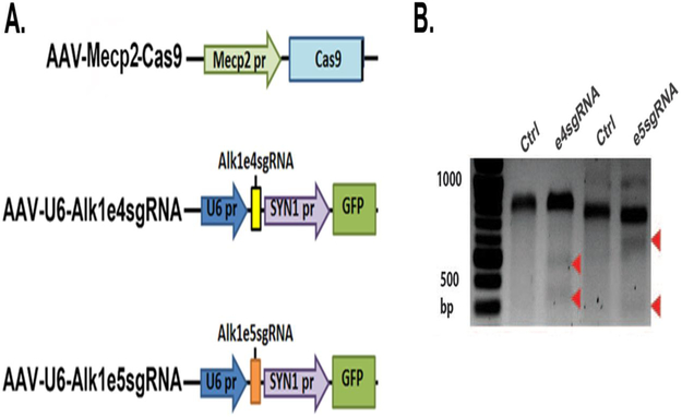
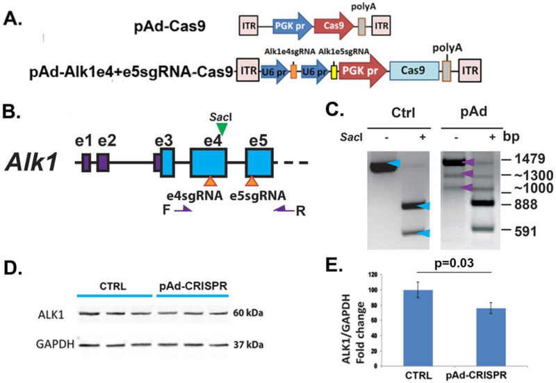
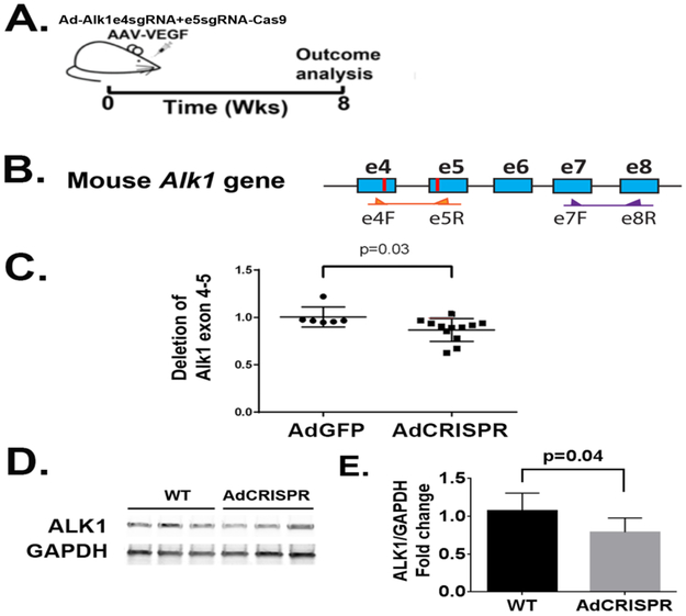
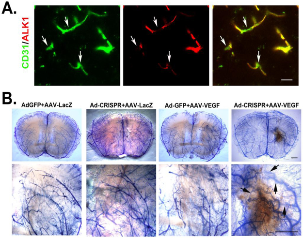
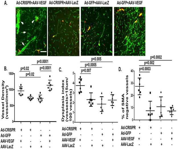
Brain arteriovenous malformation (bAVM) is an important risk factor for intracranial hemorrhage. The pathogenesis of bAVM has not been fully understood. Animal models are important tools for dissecting bAVM pathogenesis and testing new therapies. We have developed several mouse bAVM models using genetically modified mice. However, due to the body size, mouse bAVM models have some limitations. Recent studies identified somatic mutations in sporadic human bAVM. To develop a feasible tool to create sporadic bAVM in rodent and animals larger than rodent, we made tests using the CRISPR/Cas9 technique to induce somatic gene mutations in mouse brain in situ. Two sequence-specific guide RNAs (sgRNAs) targeting mouse Alk1 exons 4 and 5 were cloned into pAd-Alk1e4sgRNA + e5sgRNA-Cas9 plasmid. These sgRNAs were capable to generate mutations in Alk1 gene in mouse cell lines. After packaged into adenovirus, Ad-Alk1e4sgRNA + e5sgRNA-Cas9 was co-injected with an adeno-associated viral vector expressing vascular endothelial growth factor (AAV-VEGF) into the brains of wild-type C57BL/6J mice. Eight weeks after viral injection, bAVMs were detected in 10 of 12 mice. Compared to the control (Ad-GFP/AAV-VEGF-injected) brain, 13% of Alk1 alleles were mutated and Alk1 expression was reduced by 26% in the Ad-Alk1e4sgRNA + e5sgRNA-Cas9/AAV-VEGF-injected brains. Around the Ad-Alk1e4sgRNA + e5sgRNA-Cas9/AAV-VEGF injected site, Alk1-null endothelial cells were detected. Our data demonstrated that CRISPR/Cas9 is a feasible tool for generating bAVM model in animals.

Keywords: Alk1; Brain arteriovenous malformation; CRISPR/Cas9; Somatic gene mutation.


Conflict of interest statement

Conflict of Interest

The authors declare that they have not conflict of interest.

Figures

Figure 1.
Figure 1.. Alk1 e4sgRNA and e5sgRNA induced indel mutations in Alk1 gene in mouse cells.
A. Structure of vectors. Mecp2 pr: the promoter of a neural specific gene, Mecp2; SYN pr: the promoter of a neural specific gene, synapsin. B. Surveyor assay image. Indel mutations in Alk1 gene were detected in Neuro2A cells transfected with plasmids expressing Cas9 with e4sgRNA or e5sgRNA. For exon 4, wild-type band (Ctrl) is 761 base pairs (bp); mutant bands are 557 and 204 bps (arrows). For exon 5, wild-type band (Ctrl) is 830 bp; mutant bands are 480 and 350 bps (arrows).
Figure 2.
Figure 2.. pAd-Alk1e4sgRNA+e5sgRNA-Cas9 induced deletion mutation in Alk1 gene and reduced Alk1 expression in mouse cells.
A. Structure of pAd-Cas9 and pAd-Alk1e4sgRNA+e5sgRNA-Cas9 vectors. pr: promoter. PGK: phosphoglycerate kinase. ITR: inverted terminal repeats. B. Location of e4sgRNA and e5sgRNA sites (arrows at the bottom) and primers F and R (purple arrows) for detection of deletions between e4sgRNA and e5sgRNA sites. SacI (green arrow at the top) is a restriction enzyme that cleaves wild-type PCR product. C. Deletion mutation in pAd-Alk1e4sgRNA+e5sgRNA-Cas9 transfected Neuro2A cells. A 1479 bp (top blue and purple arrows) fragment was amplified from pAd-Cas9 trensfected cells (Ctrl). SacI cut this fragment into 591 and 888 bp (bottom two blue two arrows). Two additional bands (1300 and 1000 bps, bottom two purple arrows) containing frameshift and deletion mutations were amplified from pAd-Alk1e4sgRNA+e5sgRNA-Cas9 transfected cells (pAd). D. A representative image of western blot using protein isolated from bEnd3 cells. CTRL: pAd-Cas9 transfect bEnd.3 cells; pAd-CRISPR: pAd-Alk1e4sgRNA+e5sgRNA-Cas9 transfected bENd.3 cells. E. Quantitative analysis of Alk1 expression in bEnd3 cells. N=3.
Figure 3.
Figure 3.. Ad-Alk1e4sgRNA+e5sgRNA-Cas9 induced deletion mutation in Alk1 gene and reduced Alk1 expression in the mouse brain.
A. Design for in vivo studies. Ad-Alk1e4sgRNA+e5sgRNA-Cas9 and AAV-VEGF were co-injected into basal ganglia of mouse brain. Eight weeks after virus injection, brain vessels were casted by latex to detect AVM phenotypes. Through intra-cardiac ventricle perfusion, latex dye enters vein only in the presence of arteriovenous shunt. Due to the particle size, latex cannot pass capillaries. Dysplasia vessels were quantified on immunostained sections. Alk1 gene mutation was analyzed by qPCR. Alk1 expression was analyzed by western blot. B. Schematic of mouse Alk1 exons 4 to 8 (e4-e8). Blue boxes are exons. The red lines are e4sgRNA and e5sgRNA target sites. The primers used for detecting deletion mutations between e4sgRNA and e5sgRNA sites and to amply sequence between exons 7 and 8 are indicated by arrows. C. Quantification of deletion mutations. N=6 for Ad-GFP group and N=12 for Ad-Alk1e4sgRNA+e5RNA-Cas9 (Ad-CRISPR) group. D. A western blot image. E. Quantification of Alk1 protein expression. WT: brain tissues of mice injected with Ad-GFP (N=7). Ad-CRISPR: brain tissues of mice injected with Ad-Alk1e4sgRNA+e5sgRNA-Cas9 (N=6).
Figure 4.
Figure 4.. Ad-Alk1e4sgRNA+e5sgRNA-Cas9 silenced ALK1 expression in endothelial cells and induced bAVM in adult mouse.
A. Images show ALK1 null endothelial cells (arrows). Endothelial cells were stained by an anti-CD31 antibody (green). ALK1 expression was detected using an anti-ALK1 antibody (red). Scale bar: 20 μm. B. Representative images of latex perfused brains. Scale bar: 100 μm. The bottom pictures are zoom in images of the viral vector injected regions. Arrows indicate latex perfused veins.
Figure 5.
Figure 5.. Ad-Alk1e4sgRNA+e5sgRNA-Cas9 injected brain have more smooth muscle negative abnormal vessels.
A. Representative images of brain sections co-stained with antibodies specific to CD31 (green) and αSMA (red). Scale bar: 100 μm. B. Quantification of vessel densities (vessels/mm2). C. Quantification of dysplasia index (number of dysplasia vessels that are larger than 15 μm per 100 vessels). D. Quantification of αSMA negative vessels, Y axis shows % of vessels that were larger than 15 μm and αSMA negative. Ad-CRISPR: Ad-Alk1e4sgRNA+e5sgRNA-Cas9.

References

    1. Kim H, Su H, Weinsheimer S, Pawlikowska L, Young WL. Brain arteriovenous malformation pathogenesis: a response-to-injury paradigm. Acta Neurochir Suppl. 2011;111:83–92. - PMC - PubMed
    1. Park SO, Lee YJ, Seki T, Hong KH, Fliess N, Jiang Z, et al. ALK5- and TGFBR2-independent role of ALK1 in the pathogenesis of hereditary hemorrhagic telangiectasia type 2 (HHT2). Blood. 2008;111(2):633–42. - PMC - PubMed
    1. Allinson KR, Carvalho RL, van den Brink S, Mummery CL, Arthur HM. Generation of a floxed allele of the mouse Endoglin gene. Genesis. 2007;45(6):391–5. - PMC - PubMed
    1. Walker EJ, Su H, Shen F, Choi EJ, Oh SP, Chen G, et al. Arteriovenous malformation in the adult mouse brain resembling the human disease. Ann Neurol. 2011;69(6):954–62. - PMC - PubMed
    1. Choi EJ, Chen W, Jun K, Arthur HM, Young WL, Su H. Novel brain arteriovenous malformation mouse models for type 1 hereditary hemorrhagic telangiectasia. PLoS One. 2014;9(2):e88511. - PMC - PubMed

Publication types

Substances