. 2018 Dec 1;74(Pt 12):754-764.
doi: 10.1107/S2053230X18015273. Epub 2018 Nov 26.

Neutron and X-ray crystal structures of Lactobacillus brevis alcohol dehydrogenase reveal new insights into hydrogen-bonding pathways

Affiliations

Neutron and X-ray crystal structures of Lactobacillus brevis alcohol dehydrogenase reveal new insights into hydrogen-bonding pathways

Johannes Hermann et al. Acta Crystallogr F Struct Biol Commun. .

Abstract

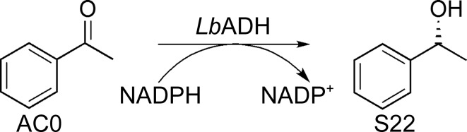
Lactobacillus brevis alcohol dehydrogenase (LbADH) is a well studied homotetrameric enzyme which catalyzes the enantioselective reduction of prochiral ketones to the corresponding secondary alcohols. LbADH is stable and enzymatically active at elevated temperatures and accepts a broad range of substrates, making it a valuable tool in industrial biocatalysis. Here, the expression, purification and crystallization of LbADH to generate large, single crystals with a volume of up to 1 mm3 suitable for neutron diffraction studies are described. Neutron diffraction data were collected from an H/D-exchanged LbADH crystal using the BIODIFF instrument at the Heinz Maier-Leibnitz Zentrum (MLZ), Garching, Germany to a resolution dmin of 2.15 Å in 16 days. This allowed the first neutron crystal structure of LbADH to be determined. The neutron structure revealed new details of the hydrogen-bonding network originating from the ion-binding site of LbADH and provided new insights into the reasons why divalent magnesium (Mg2+) or manganese (Mn2+) ions are necessary for its activity. X-ray diffraction data were obtained from the same crystal at the European Synchrotron Radiation Facility (ESRF), Grenoble, France to a resolution dmin of 1.48 Å. The high-resolution X-ray structure suggested partial occupancy of Mn2+ and Mg2+ at the ion-binding site. This is supported by the different binding affinity of Mn2+ and Mg2+ to the tetrameric structure calculated via free-energy molecular-dynamics simulations.

Keywords: Lactobacillus brevis; alcohol dehydrogenase; hydrogen-bonding network; neutron diffraction; protein crystallization; short-chain dehydrogenases/reductases.


Figures

Figure 1
Figure 1
LbADH-catalysed reduction of acetophenone (AC0) to (R)-1-phenyl­ethanol (S22).
Figure 2
Figure 2
Structural arrangement of LbADH in complex with NADP, acetophenone (AC0) and Mg2+ (PDB entry 1zk4). (a) Overall tetrameric arrangement of LbADH. The monomers are shown as differently coloured ribbon representations and the Mg2+ ion as a sphere; the NADP cofactor and AC0 substrate are highlighted as stick models (green and cyan, respectively). (b) Enlargement of the substrate and co-substrate binding region and (c) enlargement of the catalytic centre. This figure was prepared using PyMOL (v.2.1; Schrödinger).
Figure 3
Figure 3
Microscopic images of (a) the largest (∼1.0 mm3) and (b) the best neutron-diffracting His6-tagged LbADH crystal (∼0.7 mm3).
Figure 4
Figure 4
Comparison of the unit cells of the neutron (PDB entry 6h1m; orange) and X-ray (PDB entry 6h07; green) structures. Monomers A of the X-ray and neutron structures were superimposed using the Cα atoms as a reference frame. Mg2+/Mn2+ ions in the neutron structure are coloured red and those in the X-ray structure are coloured blue. This figure was prepared using PyMOL (v.2.1; Schrödinger).
Figure 5
Figure 5
R.m.s. deviation between the neutron and X-ray structures for Cα atoms. This plot was prepared with MultiSeq (Roberts et al., 2006 ▸).
Figure 6
Figure 6
Mg2+/Mn2+-binding site coordinating four water molecules and connecting two C-terminal Gln251 residues of the biological unit. A difference OMIT map (F oDF c) for both Gln251 residues, all water molecules and Mn2+/Mg2+ is contoured at 2.5σ. Red dashed lines indicate the coordination sphere for Mn2+/Mg2+. Distances are given in Å. (a) The nuclear scattering map for the neutron structure (orange; PDB entry 6h1m) shows no detectable density at the ion-binding site. Mn2+/Mg2+ was modelled in the neutron structure. (b) The electron scattering map for the X-ray structure (green; PDB entry 6h07) reveals clear density at the ion-binding site. This figure was prepared using CCP4mg (McNicholas et al., 2011 ▸).
Figure 7
Figure 7
Structural environment of the Mg2+/Mn2+ ion-binding site initiating a hydrogen-bonding pathway to Glu144. The neutron structure (orange; PDB entry 6h1m) and X-ray structure (green; PDB entry 6h07) are superimposed. A difference OMIT map (F oDF c) for Wat6 of the neutron structure is contoured at 2.5σ. Blue dashed lines mark hydrogen bonds. Red dashed lines indicate the coordination sphere for Mn2+/Mg2+. Distances are given in Å and are measured according to the neutron structure. This figure was prepared using CCP4mg (McNicholas et al., 2011 ▸).
Figure 8
Figure 8
Influence of the Mn2+/Mg2+ ion-binding site on Lys210 in the neutron structure (orange; PDB entry 6h1m) and the X-ray structure (green; PDB entry 6h07). The hydrogen bonds are marked with blue dashed lines in the neutron structure. Black dashed lines in the X-ray structure mark the distances between O atoms. Red dashed lines indicate coordinated electrostatic bonds originating from Mn2+/Mg2+. Distances are given in Å. This figure was prepared using CCP4mg (McNicholas et al., 2011 ▸).

References

    1. Adams, P. D., Afonine, P. V., Bunkóczi, G., Chen, V. B., Davis, I. W., Echols, N., Headd, J. J., Hung, L.-W., Kapral, G. J., Grosse-Kunstleve, R. W., McCoy, A. J., Moriarty, N. W., Oeffner, R., Read, R. J., Richardson, D. C., Richardson, J. S., Terwilliger, T. C. & Zwart, P. H. (2010). Acta Cryst. D66, 213–221. - PMC - PubMed
    1. Adams, P. D., Afonine, P. V., Bunkóczi, G., Chen, V. B., Echols, N., Headd, J. J., Hung, L. W., Jain, S., Kapral, G. J., Grosse Kunstleve, R. W., McCoy, A. J., Moriarty, N. W., Oeffner, R. D., Read, R. J., Richardson, D. C., Richardson, J. S., Terwilliger, T. C. & Zwart, P. H. (2011). Methods, 55, 94–106. - PMC - PubMed
    1. Afonine, P. V., Grosse-Kunstleve, R. W., Echols, N., Headd, J. J., Moriarty, N. W., Mustyakimov, M., Terwilliger, T. C., Urzhumtsev, A., Zwart, P. H. & Adams, P. D. (2012). Acta Cryst. D68, 352–367. - PMC - PubMed
    1. Allnér, O., Nilsson, L. & Villa, A. (2012). J. Chem. Theory Comput. 8, 1493–1502. - PubMed
    1. Bennett, C. H. (1976). J. Comput. Phys. 22, 245–268.

MeSH terms

Substances