. 2019 Jan 9;141(1):520-530.
doi: 10.1021/jacs.8b11373. Epub 2018 Dec 20.

Photoactivation Mechanism, Timing of Protein Secondary Structure Dynamics and Carotenoid Translocation in the Orange Carotenoid Protein

Affiliations

Photoactivation Mechanism, Timing of Protein Secondary Structure Dynamics and Carotenoid Translocation in the Orange Carotenoid Protein

Patrick E Konold et al. J Am Chem Soc. .

Abstract

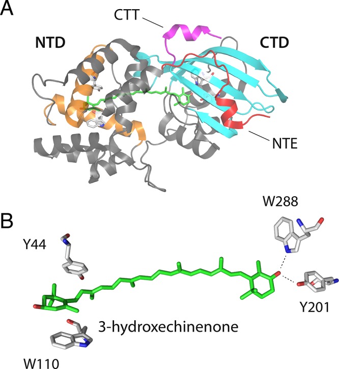
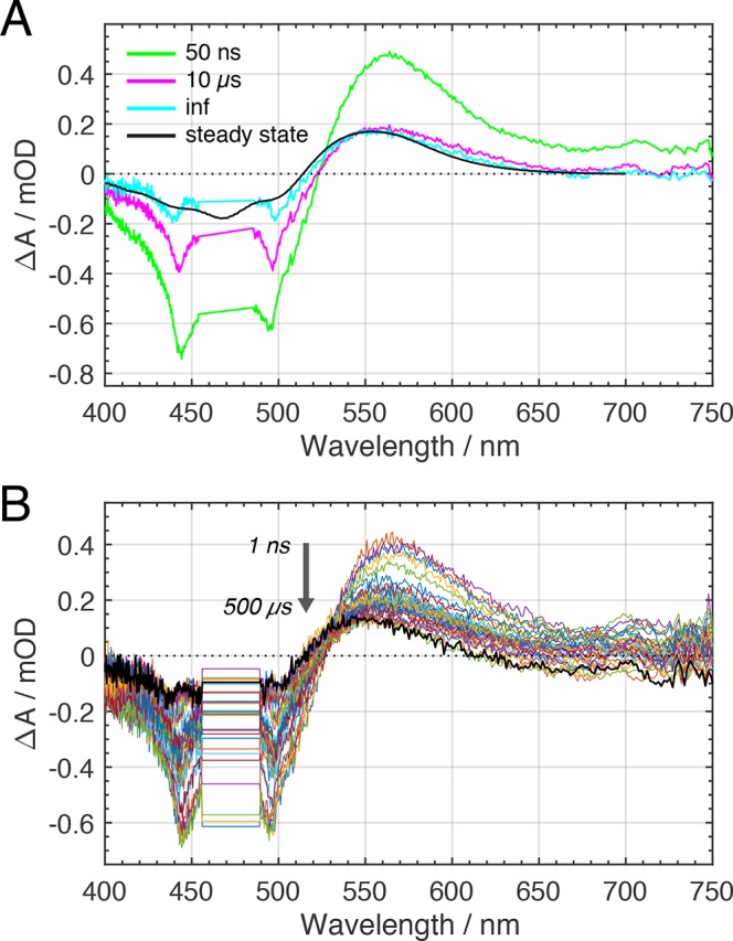
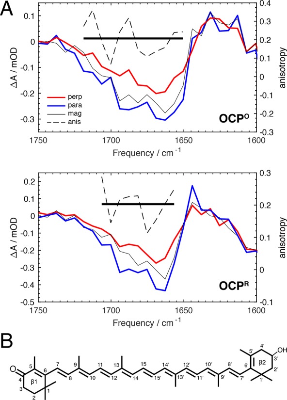
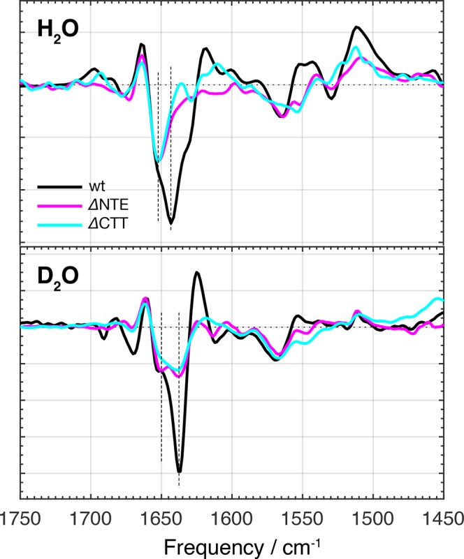
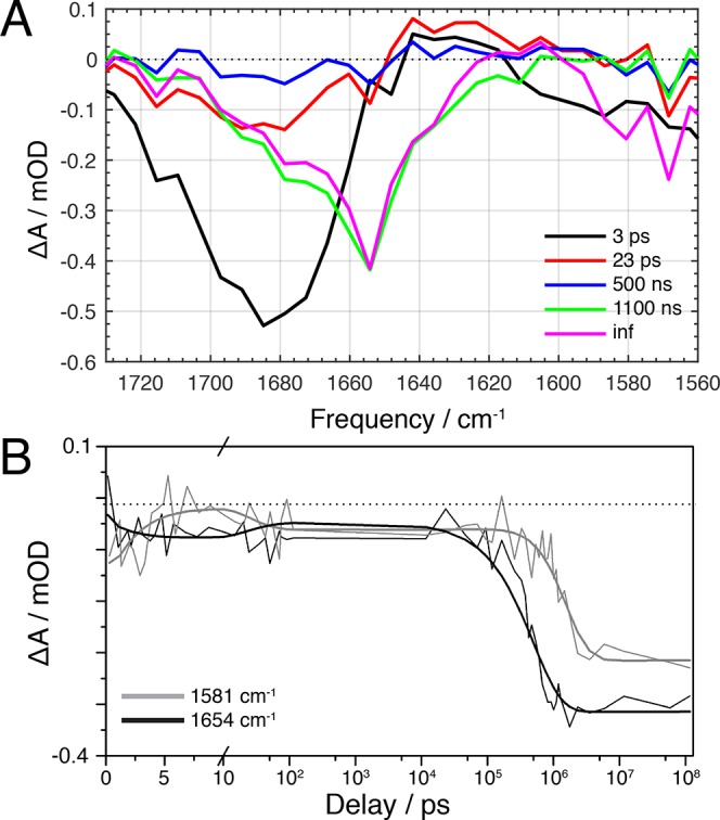
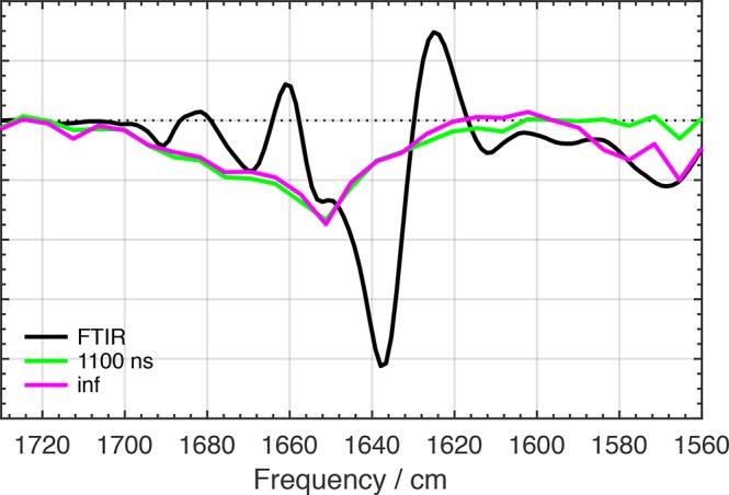
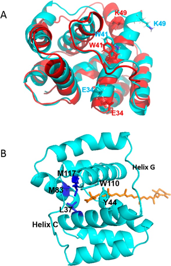
The orange carotenoid protein (OCP) is a two-domain photoactive protein that noncovalently binds an echinenone (ECN) carotenoid and mediates photoprotection in cyanobacteria. In the dark, OCP assumes an orange, inactive state known as OCPO; blue light illumination results in the red active state, known as OCPR. The OCPR state is characterized by large-scale structural changes that involve dissociation and separation of C-terminal and N-terminal domains accompanied by carotenoid translocation into the N-terminal domain. The mechanistic and dynamic-structural relations between photon absorption and formation of the OCPR state have remained largely unknown. Here, we employ a combination of time-resolved UV-visible and (polarized) mid-infrared spectroscopy to assess the electronic and structural dynamics of the carotenoid and the protein secondary structure, from femtoseconds to 0.5 ms. We identify a hereto unidentified carotenoid excited state in OCP, the so-called S* state, which we propose to play a key role in breaking conserved hydrogen-bond interactions between carotenoid and aromatic amino acids in the binding pocket. We arrive at a comprehensive reaction model where the hydrogen-bond rupture with conserved aromatic side chains at the carotenoid β1-ring in picoseconds occurs at a low yield of <1%, whereby the β1-ring retains a trans configuration with respect to the conjugated π-electron chain. This event initiates structural changes at the N-terminal domain in 1 μs, which allow the carotenoid to translocate into the N-terminal domain in 10 μs. We identified infrared signatures of helical elements that dock on the C-terminal domain β-sheet in the dark and unfold in the light to allow domain separation. These helical elements do not move within the experimental range of 0.5 ms, indicating that domain separation occurs on longer time scales, lagging carotenoid translocation by at least 2 decades of time.


Conflict of interest statement

The authors declare no competing financial interest.

Figures

Figure 1
Figure 1
(A) OCP dark state structure (OCPO) derived from X-ray crystallography (PDB: 5UI2) with mediating β-sheet (blue) and relevant terminal helical domains, NTE (red) and CTT (magenta), highlighted. (B) 3′-Hydroxyechinenone chromophore (ECN) and neighboring residues implicated in the light-induced response of OCP.
Figure 2
Figure 2
(A) Slowest EADS derived from global analysis of UV–vis TA data of OCP are overlaid with the difference (light minus dark) UV–vis absorption spectrum. (B) Raw time-gated UV–vis TA spectra from 1 ns to 500 μs. Note that the data between 454 and 485 nm have been omitted due to scatter from the excitation pulse.
Figure 3
Figure 3
(A) Mid-IR anisotropy spectra with optical excitation at 475 nm and mid-infrared probing taken at a 2 ps time delay, with parallel polarization in blue, perpendicular polarization in red, and magic angle in thin black (left vertical axis), for OCPO (upper panel) and OCPR (lower panel). The anisotropy (right vertical axis) is plotted with the dashed line, with the average indicated with the thick solid black line. (B) 3′-Hydroxyechinenone chemical structure with C6–C7 in a trans conformation, with atomic numbering included.
Figure 4
Figure 4
Light-minus-dark difference FTIR spectra of wild-type OCP (black), the δNTE mutant (magenta), and the δCTT mutant (cyan) in H2O and D2O buffers.
Figure 5
Figure 5
(A) EADS determined from global analysis of mid-IR transient absorption data. (B) Kinetic traces at 1581 (gray) and 1654 cm–1 (black), with fitting results overlaid. The 1581 cm–1 trace was expanded by a factor 2.5 to facilitate comparison with the 1654 cm–1 trace.
Figure 6
Figure 6
Light-minus-dark difference FTIR spectrum for wild-type OCP in D2O buffer (black line) with 1.1 μs EADS (green line) and nondecaying EADS (magenta line) overlaid.
Figure 7
Figure 7
Model of OCP photoactivation mechanism derived from various UV–vis and mid-IR spectroscopic experiments.
Figure 8
Figure 8
(A) Structure comparison between the NTD in OCPO (cyan) and the isolated N-terminal domain (red). The figure shows the rotation of helix C with different position of three amino acids (E34, W41, K49). (B) The structure of the N-terminal domain in the OCPO state (cyan) and ECN (orange), showing the carotenoid tunnel and the three amino acids (M117, M83, L37, in dark blue) forming the bottlenecks for carotenoid translocation. Y44 and W110, essential for photoactivation,, are placed in helices C and G, respectively. Small movement of the carotenoid may induce changes in the position of helices C and G through disruption of the π–π interactions between Y44/W110 and the ECN β2-ring. Replacement of W110 and/or Y44 by Ser abolishes photoactivation. By contrast, replacement by Phe does not hinder photoactivation, suggesting that aromatic π–π interactions with the ECN β2-ring constitute a key element.

References

    1. Horton P.; Ruban A. V.; Walters R. G. Annu. Rev. Plant Physiol. Plant Mol. Biol. 1996, 47, 655.10.1146/annurev.arplant.47.1.655. - DOI - PubMed
    1. Croce R.; van Amerongen H. Nat. Chem. Biol. 2014, 10, 492.10.1038/nchembio.1555. - DOI - PubMed
    1. Kirilovsky D.; Kerfeld C. A. Nat. Plants 2016, 2, 16180.10.1038/nplants.2016.180. - DOI - PubMed
    1. Demmigadams B. Biochim. Biophys. Acta, Bioenerg. 1990, 1020, 1.10.1016/0005-2728(90)90088-L. - DOI
    1. Horton P.; Ruban A. V.; Walters R. G. Plant Physiol. 1994, 106, 415.10.1104/pp.106.2.415. - DOI - PMC - PubMed

Publication types

Substances