

Anatomy of the Superficial Fascia System of the Breast: A Comprehensive Theory of Breast Fascial Anatomy
- PMID: 30511967
- PMCID: PMC6211786
- DOI: 10.1097/PRS.0000000000004948
Anatomy of the Superficial Fascia System of the Breast: A Comprehensive Theory of Breast Fascial Anatomy
Abstract
Background: It has been two centuries since Petrus Camper identified superficial fascia and over 175 years since Sir Astley Cooper wrote his book on the anatomy of the breast. In the 1990s, Ted Lockwood taught us the importance of the superficial fascia layers in body contouring procedures he pioneered. These descriptions, however, fail to explain the three-dimensional fascial system in the breast. The authors set out to discover and describe a theory of superficial fascia structures responsible for breast shape.
Methods: The nature of the superficial fascia system that surrounds the breast and its attachments to the chest were studied in 12 cadaver breast dissections and in clinical cases of both cosmetic and reconstructive breast procedures.
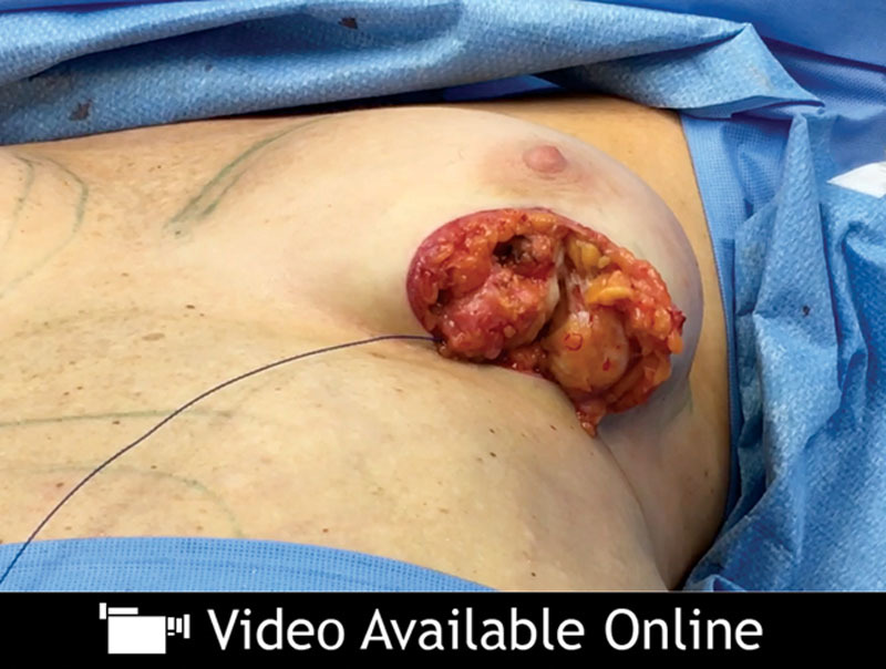
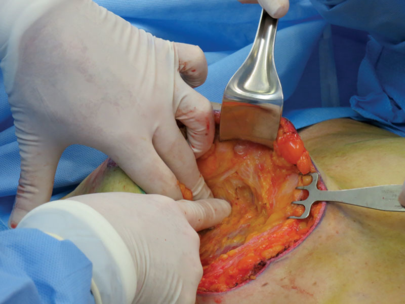
Results: The authors found a three-dimensional, closed system of fascia and fat surrounding the corpus mammae, which attaches to the skin by means of specialized vertical cutaneous ligaments, or Cooper ligaments, and which attaches to the chest wall by means of a three-dimensional zone of adherence at the breast's periphery.
Conclusions: The breast is shaped by a three-dimensional, fibrofatty fascial system. Two layers of this system surround the corpus mammae and fuse together around it, and anchor it to the chest wall in a structure we have called the circummammary ligament.
Figures


Comment in
-
Breast Anatomy: The Importance of Understanding the Superficial Fascial System for Oncoplastic Dissection.Plast Reconstr Surg. 2019 Aug;144(2):320e. doi: 10.1097/PRS.0000000000005814. Plast Reconstr Surg. 2019. PMID: 31348381 No abstract available.
-
Reply: Anatomy of the Superficial Fascia System of the Breast: A Comprehensive Theory of Breast Fascial Anatomy.Plast Reconstr Surg. 2019 Aug;144(2):320e-322e. doi: 10.1097/PRS.0000000000005815. Plast Reconstr Surg. 2019. PMID: 31348382 No abstract available.
-
Supporting a Comprehensive Theory of Breast Fascial Anatomy.Plast Reconstr Surg. 2019 Oct;144(4):706e-707e. doi: 10.1097/PRS.0000000000006032. Plast Reconstr Surg. 2019. PMID: 31568328 No abstract available.
-
Reply: Anatomy of the Superficial Fascia System of the Breast: A Comprehensive Theory of Breast Fascial Anatomy.Plast Reconstr Surg. 2019 Oct;144(4):707e-709e. doi: 10.1097/PRS.0000000000006033. Plast Reconstr Surg. 2019. PMID: 31568329 No abstract available.
-
Anatomy of the Superficial Fascia System of the Breast: A Comprehensive Theory of Breast Fascial Anatomy.Plast Reconstr Surg. 2020 Jan;145(1):193e-194e. doi: 10.1097/PRS.0000000000006329. Plast Reconstr Surg. 2020. PMID: 31881634 No abstract available.
References
-
- Cooper AP. On the Anatomy of the Breast 1840. London: Longman, Orme, Green, Brown, and Longmans;. Available at: https://jdc.jefferson.edu/cooper/60/. Accessed September 6, 2018.
-
- Stuzin JM, Baker TJ, Gordon HL. The relationship of the superficial and deep facial fascias: Relevance to rhytidectomy and aging. Plast Reconstr Surg. 1992;89:441–449; discussion 450451.. - PubMed
-
- Pessa JE. SMAS fusion zones determine the subfascial and subcutaneous anatomy of the human face: Fascial spaces, fat compartments, and models of facial aging. Aesthet Surg J. 2016;36:515–526.. - PubMed
-
- Rohrich RJ, Smith PD, Marcantonio DR, Henkel JM. The zones of adherence: Role in minimizing and preventing contour deformities in liposuction. Plast Reconstr Surg. 2001;107:1562–1569.. - PubMed
Publication types
MeSH terms
LinkOut - more resources
Full Text Sources
Other Literature Sources
