

MRI guidance technology development in a large animal model for hyperlocal analgesics delivery to the epidural space and dorsal root ganglion
- PMID: 30513305
- PMCID: PMC6317369
- DOI: 10.1016/j.jneumeth.2018.11.024
MRI guidance technology development in a large animal model for hyperlocal analgesics delivery to the epidural space and dorsal root ganglion
Abstract
Background: Development of new analgesic drugs or gene therapy vectors for spinal delivery will be facilitated by "hyperlocal" targeting of small therapeutic injectate volumes if spine imaging technology can be used that is ready for future clinical translation.
New method: This study provides methods for MRI-guided drug delivery to the periganglionic epidural space and the dorsal root ganglion (DRG) in the Yucatan swine.
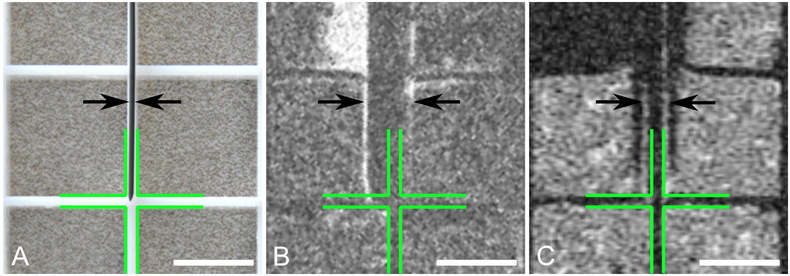
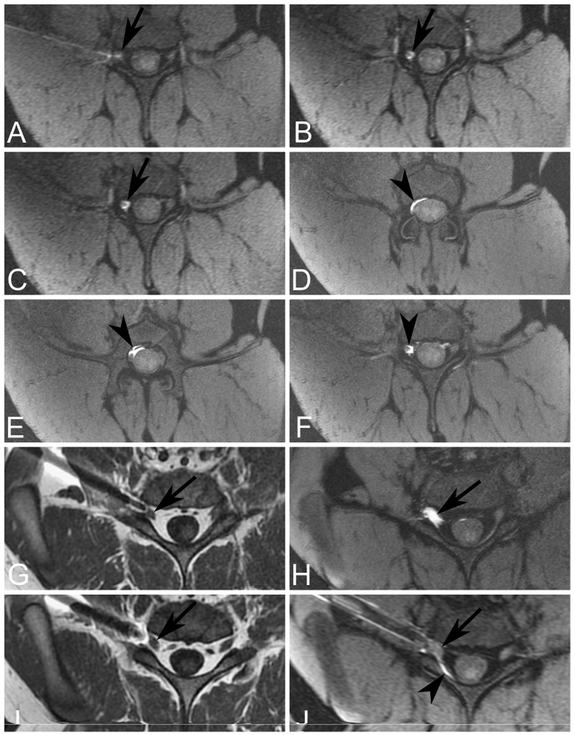
Results: Phantom studies showed artifact-corrected needle localization with frequency encoding parallel to the needle shaft, while maximizing bandwidth (125 KHz) minimized needle artifact. A custom constructed 8-12 element surface coil (phased array) wrapped over the spine in conjunction with lateral recumbent positioning achieved diagnostic quality signal to noise ratio at the depth of the DRG and afforded transforaminal access via anterolateral or posterolateral vectors, as well as interlaminar access. Swine epidural anatomy was homologous with human anatomy. Injectate containing 2% gadolinium allowed imaging of injectate volumes in increments as small as 10 microliters and discrimination of epidural flow from intraparenchymal injectate delivery into a DRG. All technical and technological elements of the procedure appear clinically translatable.
Comparison with existing methods: Computed tomographic or fluoroscopic guidance cannot directly visualize drug delivery into the DRG due to contrast medium toxicity, nor reliably identify epidural injection volumes of < 50 microliters.
Conclusions: MRI-guided hyperlocal delivery in swine provides a translatable and faithful model of future human spinal novel drug- or gene therapy vector delivery.
Keywords: Animal model; Epidural; Intraganglionic; MRI-guidance; Spinal injection.
Copyright © 2018 Elsevier B.V. All rights reserved.
Figures
Similar articles
-
Contrast flow selectivity during transforaminal lumbosacral epidural steroid injections.Pain Physician. 2008 Nov-Dec;11(6):855-61. Pain Physician. 2008. PMID: 19057631
-
Minimally invasive convection-enhanced delivery of biologics into dorsal root ganglia: validation in the pig model and prospective modeling in humans. Technical note.J Neurosurg. 2014 Oct;121(4):851-8. doi: 10.3171/2014.6.JNS132364. Epub 2014 Jul 4. J Neurosurg. 2014. PMID: 24995785 Free PMC article.
-
Correlation Between the Extent of Injectate Spread and Clinical Outcomes in Cervical Interlaminar Epidural Injection.Pain Physician. 2022 Nov;25(8):E1229-E1238. Pain Physician. 2022. PMID: 36375195
-
Fluoroscopically Guided Epidural Injections of the Cervical and Lumbar Spine.Radiographics. 2017 Mar-Apr;37(2):537-561. doi: 10.1148/rg.2017160043. Epub 2016 Dec 9. Radiographics. 2017. PMID: 27935769 Review.
-
The Anatomy, Technique, Safety, and Efficacy of Image-Guided Epidural Access.Radiol Clin North Am. 2024 Mar;62(2):199-215. doi: 10.1016/j.rcl.2023.09.006. Epub 2023 Nov 1. Radiol Clin North Am. 2024. PMID: 38272615 Review.
Cited by
-
Technical Note: A custom-designed flexible MR coil array for spine radiotherapy treatment planning.Med Phys. 2020 Jul;47(7):3143-3152. doi: 10.1002/mp.14184. Epub 2020 May 11. Med Phys. 2020. PMID: 32304237 Free PMC article.
-
Pain Treatment in the Companion Canine Model to Validate Rodent Results and Incentivize the Transition to Human Clinical Trials.Front Pharmacol. 2021 Aug 5;12:705743. doi: 10.3389/fphar.2021.705743. eCollection 2021. Front Pharmacol. 2021. PMID: 34421597 Free PMC article. Review.
References
-
- El-Yahchouchi CA, Plastaras CT, Maus TP, Carr CM, McCormick ZL, Geske JR, Smuck M, Pingree MJ, Kennedy DJ. Adverse Event Rates Associated with Transforaminal and Interlaminar Epidural Steroid Injections: A Multi-Institutional Study. Pain Med 2016; 17 (2): 239–49. Food and Drug Administration, n.d. Proceedings of the Anesthetic and Analgesic Drug Products Advisory Committee 11/24/2014. - PubMed
Publication types
MeSH terms
Substances
Grants and funding
LinkOut - more resources
Full Text Sources
Medical
