. 2018 Nov 30;19(12):3823.
doi: 10.3390/ijms19123823.

α-Melanocyte-Stimulating Hormone Attenuates Neovascularization by Inducing Nitric Oxide Deficiency via MC-Rs/PKA/NF-κB Signaling

Affiliations

α-Melanocyte-Stimulating Hormone Attenuates Neovascularization by Inducing Nitric Oxide Deficiency via MC-Rs/PKA/NF-κB Signaling

Wen-Tsan Weng et al. Int J Mol Sci. .

Abstract

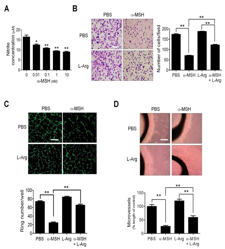
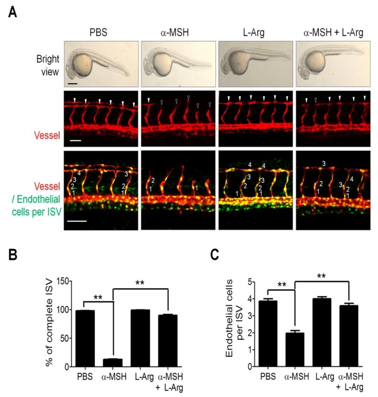
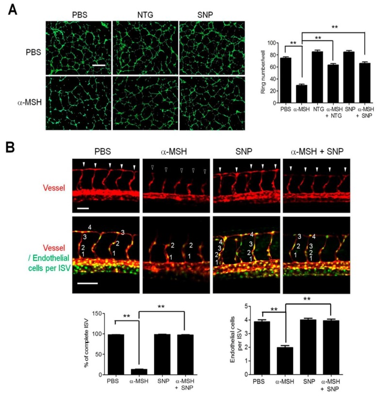
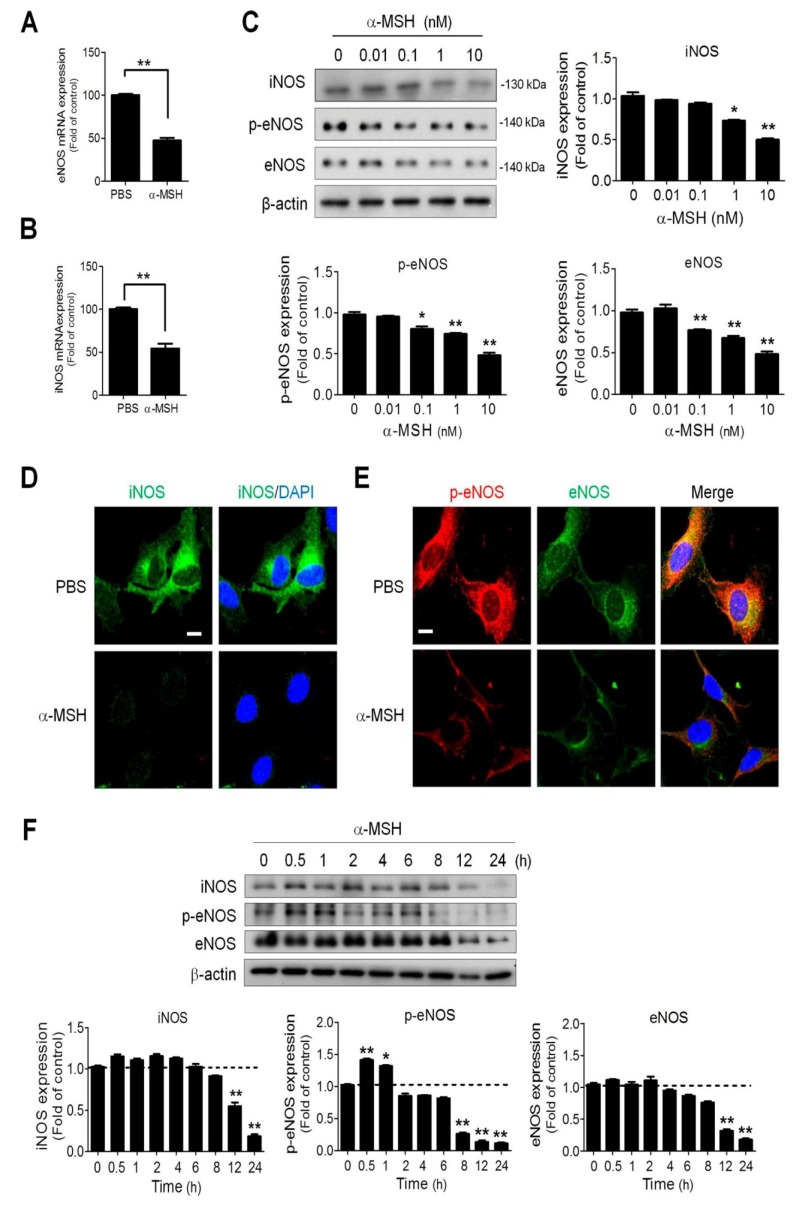
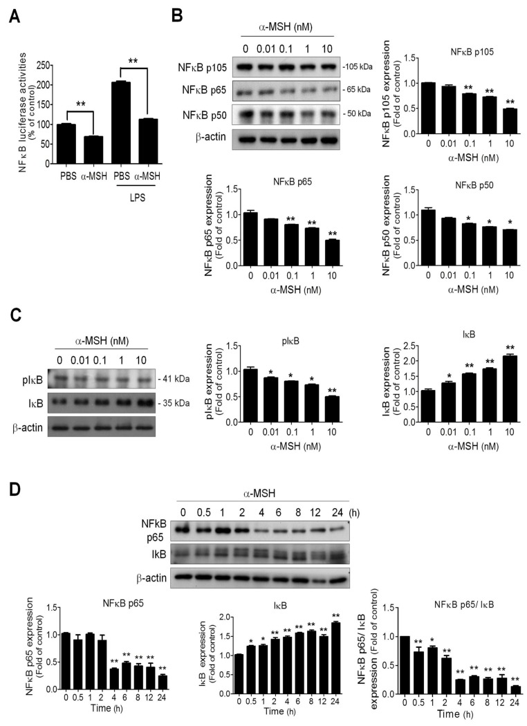
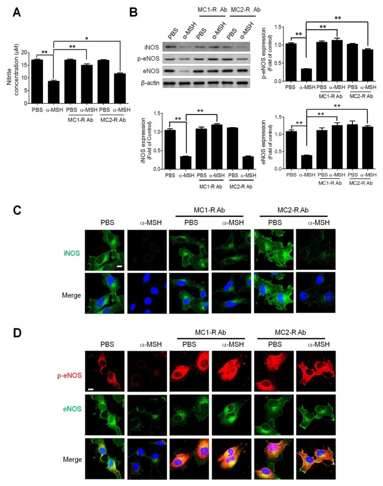
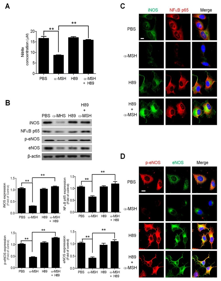
α-melanocyte-stimulating hormone (α-MSH) has been characterized as a novel angiogenesis inhibitor. The homeostasis of nitric oxide (NO) plays an important role in neovascularization. However, it remains unclear whether α-MSH mitigates angiogenesis through modulation of NO and its signaling pathway. The present study elucidated the function and mechanism of NO signaling in α-MSH-induced angiogenesis inhibition using cultured human umbilical vein endothelial cells (HUVECs), rat aorta rings, and transgenic zebrafish. By Griess reagent assay, it was found α-MSH dose-dependently reduced the NO release in HUVECs. Immunoblotting and immunofluorescence analysis revealed α-MSH potently suppressed endothelial and inducible nitric oxide synthase (eNOS/iNOS) expression, which was accompanied with inhibition of nuclear factor kappa B (NF-κB) activities. Excessive supply of NO donor l-arginine reversed the α-MSH-induced angiogenesis inhibition in vitro and in vivo. By using antibody neutralization and RNA interference, it was delineated that melanocortin-1 receptor (MC1-R) and melanocortin-2 receptor (MC2-R) participated in α-MSH-induced inhibition of NO production and NF-κB/eNOS/iNOS signaling. This was supported by pharmaceutical inhibition of protein kinase A (PKA), the downstream effector of MC-Rs signaling, using H89 abolished the α-MSH-mediated suppression of NO release and eNOS/iNOS protein level. Therefore, α-MSH exerts anti-angiogenic function by perturbing NO bioavailability and eNOS/iNOS expression in endothelial cells.

Keywords: HUVECs; melanocortin receptors (MC-Rs); nitric oxide (NO); α-melanocyte-stimulating hormone (α-MSH).


Conflict of interest statement

The authors declare no conflict of interest.

Figures

Figure 1
Figure 1
Effects of l-Arginine supply on α-MSH-induced angiogenesis inhibition in endothelial cells and aorta rings. (A) Dose-dependent effect α-Melanocyte-Stimulating Hormone (α-MSH) (0.01–10 nM for 24 h) on the nitrite levels in human umbilical vein endothelial cells (HUVECs). (B) Effect of l-Arg (5 mM) on migration in α-MSH-treated HUVECs. (C) Effect of l-Arg on tube formation in α-MSH-treated HUVECs. (D) Effect of l-Arg (5 mM) on microvessel sprouting in α-MSH-treated aortic rings. Data are expressed as mean ± SEM from triplicates. Asterisks indicate statistical significance versus control (** p < 0.001; * p < 0.005); Scale bars, 100 μm (B,C) and 2 mm (D).
Figure 2
Figure 2
Effect of l-Arginine on vascular development and endothelial recruitment in α-MSH-treated transgenic zebrafish. (A) Effect of l-Arg supply (5 mM) on vascular development and endothelial recruitment in α-MSH-treated transgenic zebrafish. Representative confocal images of phenotype and vascular growth of ISV in Tg(kdrl:mCherryci5;fli1a:negfpy5) double transgenic zebrafish embryos. The l-Arg-treated embryos displayed minor responses to the α-MSH inhibition of vessel formation (red fluorescence) and endothelial cell recruitment (green fluorescence in cell nuclei). Solid and hollow arrowheads indicate complete and incomplete vessel formation in ISV. (B) Quantification analysis of the ISV completion rate in ISV of α-MSH-treated transgenic zebrafish. (C) Quantification analysis of the number of endothelial cells per ISV of α-MSH-treated transgenic zebrafish. Data are expressed as mean ± SEM from 30 ISVs in at least 3 embryos. Asterisks indicated statistical significance versus control (** p < 0.001); Scale bars in the upper, middle, and lower panels were 50 μm, 50 μm and 100 μm, respectively.
Figure 3
Figure 3
Effect of NO/sGC modulators on the α-MSH-induced angiogenesis in vitro and in vivo. (A) Effect of synthetic NO donors (NTG and SNP; 10 μM) on α-MSH-induced inhibition of tube formation in HUVECs. Data are expressed as mean ± SEM from triplicates. Scale bars, 100 μm. (B) Effect of SNP supply on vascular development and endothelial recruitment in α-MSH-treated transgenic zebrafish. Confocal images of vessels (red fluorescence) and endothelial cells (green fluorescence) in ISV in the α-MSH-stressed zebrafish embryos after SNP treatment (10 μM). The ISV morphogenesis and endothelial cell number in each ISV were expressed as mean ± SEM calculated from 30 ISVs in at least 3 embryos. Solid and hollow arrowheads indicate complete and incomplete vessel formation in ISV. Scale bars in the upper and lower panels were 50 and 100 μm, respectively. Data were expressed as mean ± SEM from triplicate experiments. Asterisks indicate statistical significance versus control (** p < 0.001).
Figure 4
Figure 4
Dose- and time-dependent effect of α-MSH on eNOS phosphorylation and eNOS/iNOS expression in HUVECs. The inhibitory effects of α-MSH on (A) eNOS and (B) iNOS mRNA expression in HUVECs. (C) Dose effect of α-MSH on eNOS phosphorylation and eNOS/iNOS expression in HUVECs. After treatment with α-MSH (0.01–10 nM) for 24 h, the protein extracts of HUVECs were subjected to immunoblot analysis using antibodies against iNOS, phospho-Ser1177-eNOS and eNOS. (D) Immunofluorescence analysis of α-MSH treatment (10 nM for 24 h) on iNOS expression (green) in HUVECs. (E) Immunofluorescence analysis of α-MSH treatment (10 nM for 24 h) on eNOS phosphorylation (phospho-Ser1177-eNOS in red) and eNOS (in green) expression in HUVECs. The cell nuclei were stained with DAPI (blue). (F) Time-dependent effect of α-MSH on eNOS phosphorylation and eNOS/iNOS expression in HUVECs. After treatment with α-MSH (10 nM) for different intervals, the protein extracts of HUVECs were subjected to immunoblot analysis. Data were expressed as mean ± SEM calculated from triplicates. Quantification indicated mean fold change compared with the control after the expression levels were normalized to β-actin levels. Scale bars, 100 μm; * p < 0.05 and ** p < 0.01 compared with the control groups.
Figure 5
Figure 5
Effect of α-MSH on NF-κB activities and signaling in HUVECs. (A) Effect of α-MSH (10 nM for 24 h) on basal and LPS-stimulated NF-κB-driven luciferase activities in HUVECs. (B) Dose effect of α-MSH (0.01–10 nM for 24 h) on NF-κB subunits p105, p65, p50 expression in HUVECs. (C) Dose effect (0.01–10 nM for 24 h) of α-MSH on phosphorylated IκB and IκB expression in HUVECs. (D) Time-dependent effect of α-MSH (10 nM) on NF-κB p65 and IκB expression in HUVECs. After treatment with α-MSH (10 nM) for different intervals, the protein extracts of HUVECs were subjected to immunoblot analysis. Data were expressed as mean ± SEM from triplicates. Quantification indicated mean fold change compared with the control after the expression levels were normalized to β-actin levels. Scale bars, 100 μm. * p < 0.05 and ** p < 0.01 compared with the control groups.
Figure 6
Figure 6
Effect of MC1-R and MC2-R neutralization on NO release and iNOS /eNOS expression in HUVECs. (A) Effect of MC1-R and MC2-R neutralization on nitrite production in HUVECs after α-MSH treatment (10 nM for 24 h). (B) Immunoblot analysis of the effect of MC1-R and MC2-R neutralization on phospho-Ser1177 eNOS phosphorylation and eNOS/iNOS expression in α-MSH-treated HUVECs. (C) Immunofluorescence analysis of the effect of MC1-R and MC2-R neutralization on iNOS expression (green) in α-MSH-treated HUVECs. (D) Immunofluorescence analysis of the effect of MC1-R and MC2-R neutralization on phospho-Ser1177-eNOS (red) and eNOS (green) expression. Cell nuclei were counterstained with DAPI (blue). Data are expressed as mean ± SEM calculated from triplicates. Quantification indicated mean fold change compared with the control after the expression levels were normalized to β-actin levels.Scale bars, 100 μm. * p < 0.05 and ** p < 0.01 compared with the control groups.
Figure 7
Figure 7
Effects of PKA antagonist H89 on NO homeostasis and eNOS/iNOS expression in α-MSH-treated HUVECs. (A) Effect of H89 (10 μM) on nitrite production in HUVECs. (B) Immunoblot analysis of the effect of H89 on eNOS, phospho-Ser1177-eNOS, iNOS, and NF-κB p65 expression in α-MSH-treated HUVECs. (C) Immunofluorescence analysis of the effect of H89 on iNOS (green) and NF-κB p65 (red) level in α-MSH-treated HUVECs. (D) Immunofluorescence analysis of the effect of H89 on phospho-Ser1177-eNOS (red) and eNOS (green) expression in α-MSH-treated HUVECs. Cell nuclei were counterstained with DAPI (blue). Scale bars, 100 μm. Data are expressed as mean ± SEM from triplicates. Quantification indicated mean fold change compared with the control after the expression levels were normalized to β-actin levels. Scale bars, 100 μm. ** p < 0.01 compared with the control groups.

References

    1. Brzoska T., Luger T.A., Maaser C., Abels C., Bohm M. α-melanocyte-stimulating hormone and related tripeptides: Biochemistry, antiinflammatory and protective effects in vitro and in vivo, and future perspectives for the treatment of immune-mediated inflammatory diseases. Endocr. Rev. 2008;29:581–602. doi: 10.1210/er.2007-0027. - DOI - PubMed
    1. Oh C.T., Lee D., Koo K., Lee J., Yoon H.S., Choi Y.M., Kwon T.R., Kim B.J. Superoxide dismutase 1 inhibits α-melanocyte stimulating hormone and ultraviolet B-induced melanogenesis in murine skin. Ann. Dermatol. 2014;26:681–687. doi: 10.5021/ad.2014.26.6.681. - DOI - PMC - PubMed
    1. Cyr N.E., Steger J.S., Toorie A.M., Yang J.Z., Stuart R., Nillni E.A. Central Sirt1 regulates body weight and energy expenditure along with the POMC-derived peptide α-MSH and the processing enzyme CPE production in diet-induced obese male rats. Endocrinology. 2015;156:961–974. doi: 10.1210/en.2014-1970. - DOI - PMC - PubMed
    1. Murray C.J., Lopez A.D. Measuring the global burden of disease. N. Engl. J. Med. 2013;369:448–457. doi: 10.1056/NEJMra1201534. - DOI - PubMed
    1. Nabel E.G., Braunwald E. A tale of coronary artery disease and myocardial infarction. N. Engl. J. Med. 2012;366:54–63. doi: 10.1056/NEJMra1112570. - DOI - PubMed

MeSH terms

LinkOut - more resources