. 2018 Nov 30;10(12):479.
doi: 10.3390/cancers10120479.

Helicobacter pylori Infection-Induced Hepatoma-Derived Growth Factor Regulates the Differentiation of Human Mesenchymal Stem Cells to Myofibroblast-Like Cells

Affiliations

Helicobacter pylori Infection-Induced Hepatoma-Derived Growth Factor Regulates the Differentiation of Human Mesenchymal Stem Cells to Myofibroblast-Like Cells

Chung-Jung Liu et al. Cancers (Basel). .

Abstract

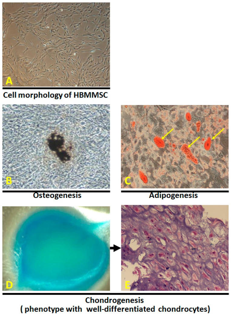
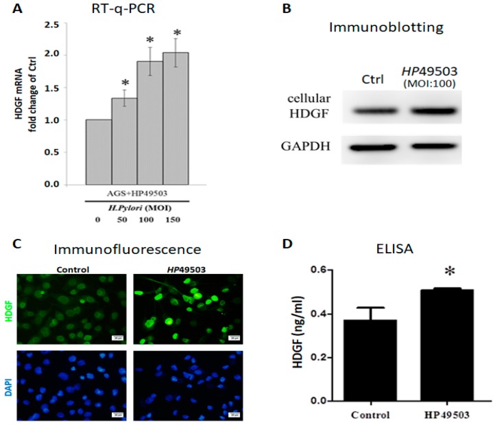
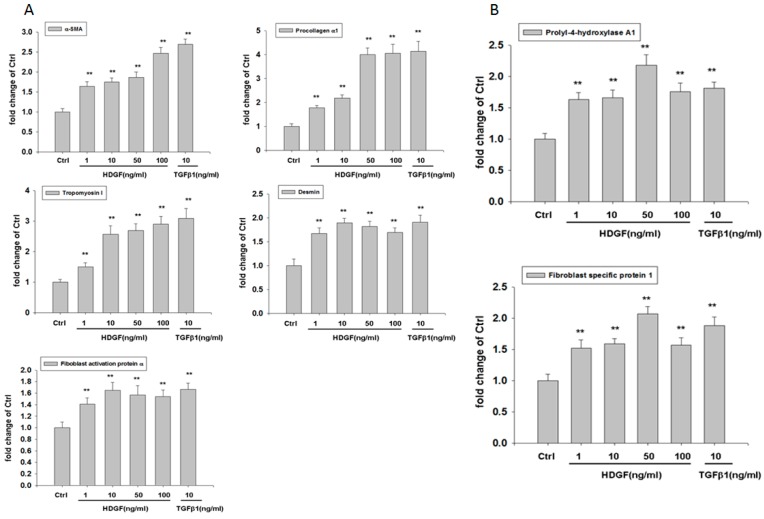
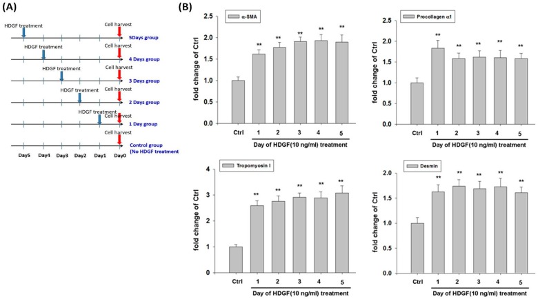
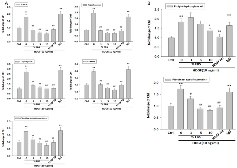
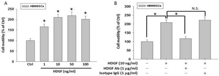
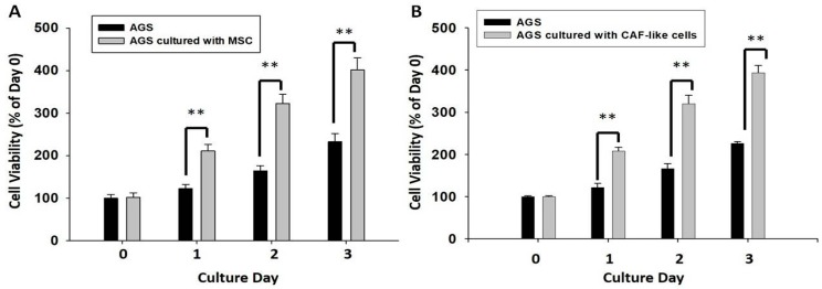
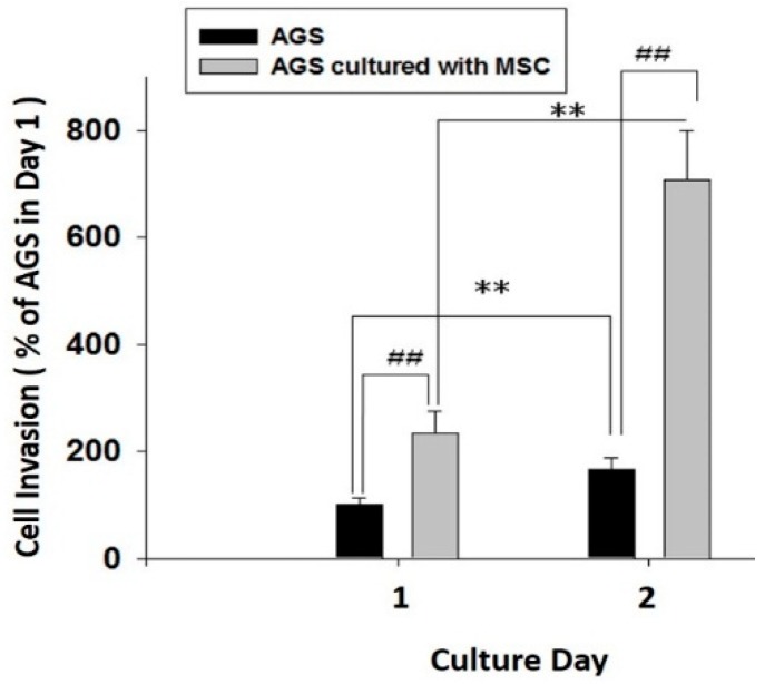
Hepatoma-derived growth factor (HDGF) plays a critical role in tumor cell proliferation, anti-apoptosis, VEGF expression, lymph node metastasis and poor prognosis in human gastric cancer. Gastric cancer, as one of the most prevalent cancers worldwide, is the second leading cause of cancer-related mortality in the world for the prognosis of gastric cancer is generally poor, especially in patients with advanced stage. Helicobacter pylori (H. pylori) infection causes the chronic inflammation of stomach as well as the development of gastric cancer, with a three to six-fold increased risk of gastric cancer. Carcinoma-associated fibroblasts (CAFs) are myofibroblasts in tumor microenvironment, which possess various abilities to promote the progression of cancer by stimulating neoangiogenesis, proliferation, migration, invasion and therapy resistance of tumor cell. Mesenchymal stem cells (MSCs) are reported to promote tumor malignance through differentiation of MSCs toward CAFs. In the present study, we demonstrated that H. pylori infection promotes HDGF expression in human gastric cancer cells. HBMMSCs treated with HDGF assume properties of CAF-like myofibroblastic phenotypes, including expression of myofibroblast markers (α-smooth muscle actin (α-SMA), procollagen α1, tropomyoson I, desmin, fibroblast activation protein (FAP)), and fibroblast markers (prolyl-4-hydroxylase A1 (PHA1) and fibroblast specific protein-1 (FSP-1)/S100A4). HDGF recruits HBMMSCs, and then HBMMSCs further contributes to cell survival and invasive motility in human gastric cancer cells. Treatment of HDGF neutralizing antibody (HDGF-NAb) and serum significantly inhibit HDGF-regulated differentiation and recruitment of HBMMSCs. These findings suggest that HDGF might play a critical role in gastric cancer progress through stimulation of HBMMSCs differentiation to myofibroblast-like cells.

Keywords: Helicobacter pylori; carcinoma-associated fibroblast; hepatoma-derived growth factor; mesenchymal stem cells; myofibroblast.


Conflict of interest statement

The authors declare no conflict of interest.

Figures

Figure 1
Figure 1
The morphology of HBMMSCs was showed by light microscopy (A). Osteogenic, chondrogenic, and adipogenic differentiation from HBMMSCs. Osteogenic differentiation from HBMMSCs was evidenced by cells morphology after 21 days of induction. Formation of mineralized matrix was shown by von Kossa staining (B, shown at original magnification ×200). Adipocytic differentiation from HBMMSCs was evidenced by the formation of lipid vacuoles by oil-red O staining (C, shown in phase-contrast photograph at original magnification ×200 and). Chondrogenic differentiation from HBMMSCs was evidenced by alcian blue staining (D), and by H&E staining for histological analysis (E).
Figure 2
Figure 2
Human AGS cells with HP infection expresses high HDGF level. HDGF mRNA in AGS cells with HP49503 infection for 24 h was analyzed by RT-q-PCR (A). HDGF protein from AGS cells infected with HP49503 for 24 h was detected by immunoblotting assay. GAPDH was used as an internal control (B). Immunofluorescent staining was used to detect the expression of HDGF in AGS cells with HP49503 infection. Scale bar = 20 μm. (C). Secreted HDGF from AGS cells with HP49503 infection was measured by ELISA assay (D). * p < 0.05 versus control (mean ± SD, n = 3).
Figure 3
Figure 3
HDGF stimulates expression of myofibroblast (A) and fibroblast (B) markers in HBMMSCs in various concentrations. HBMMSCs were treated with HDGF (1, 10, 50, 100 ng/ml) for 24 hours. The effect of HDGF on marker expression of myofibroblast and fibroblast were measured by RT q-PCR. TGFβ1 (10 ng/ml) treatment as positive control. ** p < 0.01 versus control (mean ± SD, n = 3).
Figure 4
Figure 4
(A) A schematic representation showed the procedure of HDGF treatment in HBMMSCs culture. HBMMSC were treated with HDGF at various time points in the absence of serum. HBMMSC then were harvested at the same day. (B) HDGF stimulates expression of myofibroblast markers in HBMMSCs. HBMMSCs were treated with HDGF 10 ng/ml) for various periods (1, 2, 3, 4 or 5 days), respectively. The effect of HDGF on expression of myofibroblast markers was measured by RT q-PCR. ** p < 0.01 versus control (mean ± SD, n = 3).
Figure 5
Figure 5
Serum and HDGF neutralizing antibody inhibits the expression of myofibroblast and fibroblast markers. HBMMSCs were treated with various concentration of FBS (0, 1, 5, 10%), or HDGF neutralizing antibody (HDGF-NAb; 1 μg/ml) in the presence of HDGF (10 ng/ml). After 24 hours, the marker expression of myofibroblast (A) and fibroblast (B) was measured by RT q-PCR. * p < 0.05; ** p < 0.01 versus control (line 1); # p < 0.05; ## p < 0.01 versus only HDGF treatment (line 2) (mean ± SD, n = 3).
Figure 6
Figure 6
(A) HDGF induces HBMSCs motility at various concentrations. HBMMSCs were treated with HDGF (1, 10, 50, 100 ng/ml) for 24h. The effect of HDGF on motility capacity of HBMMSCs was measured by motility assay. (B) HDGF neutralizing antibody inhibits HDGF-mediated HBMMSCs recruitment. HBMMSCs treated with HDGF neutralizing antibody (HDGF-NAb; 1 μg/ml) in the presence or absence of HDGF (10 ng/ml) were analyzed for the inhibitory effect of HDGF-Nab on HDGF-mediated recruitment motility. * p < 0.01 versus control (line 1); * p < 0.01 versus only HDGF treatment (line 2) (mean ± SD, n = 3).
Figure 7
Figure 7
HBMMSCs (A) or CAF-like cells (B) enhance survival in human gastric cancer cells. HBMMSCs or CAF-like-cells were co-cultured with human AGS gastric cancer cells for various periods (1, 2, 3 days). The effect of HBMMSCs or CAF-like cells on cellular survival of human gastric cancer cells was measured. The responses to HBMMSCs or CAF-like cells were measured by MTT assay. ** p < 0.01 versus only AGS cells (mean ± SD, n = 3).
Figure 8
Figure 8
HBMMSCs enhances invasive motility in human gastric cancer cells. HBMMSCs and human AGS gastric cancer cells were cultured for 24 and 48 h, respectively. The effect of HBMMSCs on cellular motility of human gastric cancer cells were measured. The responses to HBMMSCs were measured by invasive motility assay. ** p < 0.01, only AGS cells in day 2 versus only AGS cells in day 1; ## p < 0.01, AGS cells cultured with MSC versus only AGS cells (mean ± SD, n = 3).

References

    1. Shiga K., Hara M., Nagasaki T., Sato T., Takahashi H., Takeyama H. Cancer-Associated Fibroblasts: Their Characteristics and Their Roles in Tumor Growth. Cancers. 2015;7:2443–2458. doi: 10.3390/cancers7040902. - DOI - PMC - PubMed
    1. Kharaishvili G., Simkova D., Bouchalova K., Gachechiladze M., Narsia N., Bouchal J. The role of cancer-associated fibroblasts, solid stress and other microenvironmental factors in tumor progression and therapy resistance. Cancer Cell Int. 2014;14:41. doi: 10.1186/1475-2867-14-41. - DOI - PMC - PubMed
    1. Tang D., Gao J., Wang S., Ye N., Chong Y., Huang Y., Wang J., Li B., Yin W., Wang D. Cancer-associated fibroblasts promote angiogenesis in gastric cancer through galectin-1 expression. Tumour Biol. 2016;37:1889–1899. doi: 10.1007/s13277-015-3942-9. - DOI - PubMed
    1. Kurashige J., Mima K., Sawada G., Takahashi Y., Eguchi H., Sugimachi K., Mori M., Yanagihara K., Yashiro M., Hirakawa K., et al. Epigenetic modulation and repression of miR-200b by cancer-associated fibroblasts contribute to cancer invasion and peritoneal dissemination in gastric cancer. Carcinogenesis. 2015;36:133–141. doi: 10.1093/carcin/bgu232. - DOI - PubMed
    1. Sun C., Fukui H., Hara K., Zhang X., Kitayama Y., Eda H., Tomita T., Oshima T., Kikuchi S., Watari J., et al. FGF9 from cancer-associated fibroblasts is a possible mediator of invasion and anti-apoptosis of gastric cancer cells. BMC Cancer. 2015;15:333. doi: 10.1186/s12885-015-1353-3. - DOI - PMC - PubMed