Review
. 2018 Dec 3;19(12):3850.
doi: 10.3390/ijms19123850.

Genetic Hierarchy of Acute Myeloid Leukemia: From Clonal Hematopoiesis to Molecular Residual Disease

Affiliations
Review

Genetic Hierarchy of Acute Myeloid Leukemia: From Clonal Hematopoiesis to Molecular Residual Disease

Jean-Alain Martignoles et al. Int J Mol Sci. .

Abstract

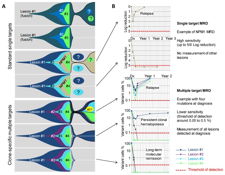
Recent advances in the field of cancer genome analysis revolutionized the picture we have of acute myeloid leukemia (AML). Pan-genomic studies, using either single nucleotide polymorphism arrays or whole genome/exome next generation sequencing, uncovered alterations in dozens of new genes or pathways, intimately connected with the development of leukemia. From a simple two-hit model in the late nineties, we are now building clonal stories that involve multiple unexpected cellular functions, leading to full-blown AML. In this review, we will address several seminal concepts that result from these new findings. We will describe the genetic landscape of AML, the association and order of events that define multiple sub-entities, both in terms of pathogenesis and in terms of clinical practice. Finally, we will discuss the use of this knowledge in the settings of new strategies for the evaluation of measurable residual diseases (MRD), using clone-specific multiple molecular targets.

Keywords: acute myeloid leukemia; clonal hematopoiesis; genetic hierarchy; molecular residual disease.


Conflict of interest statement

The authors declare no conflict of interest.

Figures

Figure 1
Figure 1
Associations and orders of acquired lesions in de novo, secondary, and therapy-related AMLs. (A) A schematic view of the multiple lesions underlying leukemogenesis and AML ontology, according to the classification in de-novo-type, secondary-type, and TP53-type AMLs—central lesions are shared by all types of AMLs, whereas outlying ones are either specific to or enriched in the type of AML indicated by the nearest arrow. Only the most frequent lesions are indicated in each category. (B) Fish diagrams of representative (but not exhaustive) clonal AML hierarchies in de novo AMLs. Grey areas show normal hematopoietic stem cells and their progeny. The onset of a clone is achieved through the acquisition of a genetic lesion (first lesion) and the subsequent expansion of mutant cells (colored cells and areas), as indicated in the top panel. Subsequent events will shape the clones with time and lead to AML. Lesions are color-coded, as indicated in panel A. (C) Fish diagrams showing representative (but not exhaustive) clonal AML hierarchies in secondary AMLs (s-AML) and therapy-related AMLs (t-AML), as in B.
Figure 2
Figure 2
Possible outcomes in AML. Fish diagrams showing clonal evolution in the four possible AML outcomes. Refractoriness and early relapse are diagnosed using cytomorphology and are characterized by the early persistence of most, if not all, lesions, found at the diagnosis. Later relapse occurs after a period of complete remission, where no excess blast is seen, but where most lesions persist at low levels. A peculiar condition is represented by the persistence of clonal hematopoiesis in patients who are in long-lasting remission but retain a founder clone with the most frequent DNMT3A mutations. Finally, patients are cured when all mutant cells are cleared from the bone marrow. In these last three outcomes, cytometric and molecular measurable residual diseases (MRD) evaluations are critical for patient monitoring. #1, #2, #3 etc. indicate the successive genetic lesions.
Figure 3
Figure 3
Molecular MRD using single and multiple targets to track AML clones. (A) Fish diagrams showing clonal evolution in AMLs with various combinations of lesions and outcomes. MRD Target lesions are indicated by contoured areas in the right part (post-diagnosis) of the diagrams. Black contours indicate lesions that are found MRD-positive, and white contours and dotted lines indicate lesions that become progressively not detectable. Circles with question marks indicate lesions that are not detected by the MRD test. (B) Simulations of MRD monitoring of five cases from (A) using a single target MRD test (upper panels) or a clone-specific, multi-target MRD approach (lower panels). Red lines indicate the theoretical sensitivity of the assay. #1, #2, #3 etc. indicate the successive genetic lesions.

References

    1. Arber D.A., Orazi A., Hasserjian R., Thiele J., Borowitz M.J., le Beau M.M., Bloomfield C.D., Cazzola M., Vardiman J.W. The 2016 revision to the World Health Organization classification of myeloid neoplasms and acute leukemia. Blood. 2016;127:2391–2405. doi: 10.1182/blood-2016-03-643544. - DOI - PubMed
    1. Papaemmanuil E., Gerstung M., Bullinger L., Gaidzik V.I., Paschka P., Roberts N.D., Potter N.E., Heuser M., Thol F., Bolli N., et al. Genomic classification and prognosis in acute myeloid leukemia. N. Engl. J. Med. 2016;374:2209–2221. doi: 10.1056/NEJMoa1516192. - DOI - PMC - PubMed
    1. Lindsley R.C., Mar B.G., Mazzola E., Grauman P.V., Shareef S., Allen S.L., Pigneux A., Wetzler M., Stuart R.K., Erba H.P., et al. Acute myeloid leukemia ontogeny is defined by distinct somatic mutations. Blood. 2015;125:1367–1376. doi: 10.1182/blood-2014-11-610543. - DOI - PMC - PubMed
    1. Welch J.S., Ley T.J., Link D.C., Miller C.A., Larson D.E., Koboldt D.C., Wartman L.D., Lamprecht T.L., Liu F., Xia J., et al. The origin and evolution of mutations in acute myeloid leukemia. Cell. 2012;150:264–278. doi: 10.1016/j.cell.2012.06.023. - DOI - PMC - PubMed
    1. Cancer Genome Atlas Research Network. Ley T.J., Miller C., Ding L., Raphael B.J., Mungall A.J., Robertson A.G., Hoadley K., Triche T.J., Laird P.W., et al. Genomic and epigenomic landscapes of adult de novo acute myeloid leukemia. N. Engl. J. Med. 2013;368:2059–2074. doi: 10.1056/NEJMoa1301689. - DOI - PMC - PubMed

MeSH terms