Case Reports
. 2018 Dec 4;14(1):382.
doi: 10.1186/s12917-018-1710-y.

Clinical response to isotretinoin and interferon-α of two dogs with cutaneous epitheliotropic T-cell lymphoma: a case report

Affiliations
Case Reports

Clinical response to isotretinoin and interferon-α of two dogs with cutaneous epitheliotropic T-cell lymphoma: a case report

Ga-Won Lee et al. BMC Vet Res. .

Abstract

Background: There is no specific therapy for cutaneous epitheliotropic T-cell lymphoma (CETL). The administration of retinoids in conjunction with interferon-α (IFN-α) in CETL has not been reported in dogs.

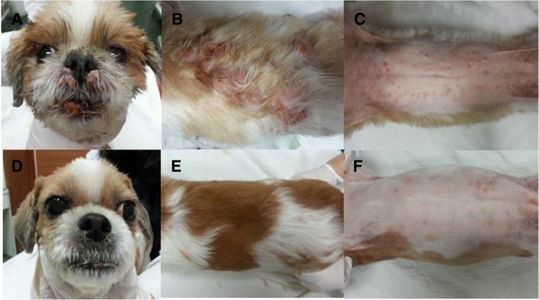
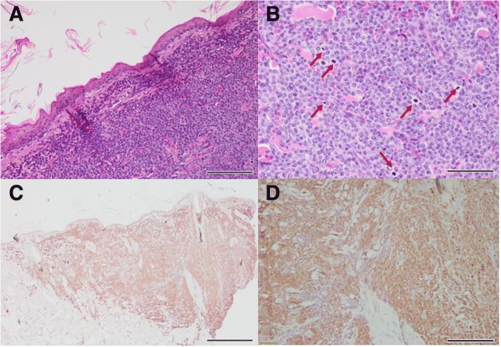
Case presentation: Two dogs (Shih tzu and Miniature pinscher) presented with multiple nodular skin lesions. Histopathological examination revealed diffuse infiltrations of lymphocytes in the epidermis and dermis, with a CD3-positive immunophenotypic profile. Based on the clinical and histopathological examination, CETL was diagnosed. Both dogs were treated with isotretinoin in combination with IFN-α and showed clinical improvement with complete or partial remission. The disease in these dogs was well-controlled for more than 264 days of overall median survival time without any additional clinical signs after initiation of the treatment. In both the cases, the dogs were followed up for 27 months, and 10 months without any evidence of recurrence or metastasis, respectively.

Conclusions: We describe the clinical efficacy of isotretinoin combined with IFN-α in 2 dogs with CETL. Long-term management with isotretinoin combined with IFN-α was effective in treating CETL in these cases.

Keywords: Canine; Cutaneous T-cell lymphoma; Interferon-α; Retinoids.


Conflict of interest statement

Ethics approval and consent to participate

Not applicable.

Consent to publication

Written consent was obtained from the present owners of the dogs for publication of this case report and any accompanying images.

Competing interests

The authors declare that they have no competing interests.

Publisher’s Note

Springer Nature remains neutral with regard to jurisdictional claims in published maps and institutional affiliations.

Figures

Fig. 1
Fig. 1
General appearance of epitheliotropic lymphoma in a Shih tzu dog (case 1). Multiple erythematous nodules were marked on the face (a) and the dorsum (b), and multiple papules detected on the ventral skin lesion (c). Three months after initiation of isotretinoin in conjunction with IFN-α shows complete disappearance of nodules and complete remission (d, e, and f)
Fig. 2
Fig. 2
Histopathology of nodules of dorsum and trunk of case 1. Note the tropism of neoplastic lymphocytes in the epidermis (a; bar = 20 μm, H&E stain) and apocrine sweat glands (b: bar = 100 μm, H&E stain), with formation of Pautrier’s microabscesses (arrows), indicating mycosis fungoides. Immunohistochemical results show numerous CD3 positive (c: CD3, bar = 200 μm) & CD79a negative cells (d: CD79a, bar = 200 μm), indicating T-cell epitheliotropic lymphoma
Fig. 3
Fig. 3
General appearance of epitheliotropic lymphoma in a Miniature pinscher dog (case 2). Generalized ulcerative nodules, characteristic of epitheliotropic lymphoma, were detected at the time of diagnosis (a, c, e, and g) and 2 weeks after administration of isotretinoin with IFN-α (b, d, f, and h). Skin lesions remarkably improved 2 weeks after multimodality therapy. The improvement of clinical features was well maintained up to 2 months after diagnosis
Fig. 4
Fig. 4
Histopathology of nodules of dorsum and trunk of case 2. Epidermal and dermal infiltration of lymphoid cells with large nuclei and high mitotic activity (red arrows) are shown in A & B (a: bar = 100 μm, b: bar = 50 μm; H&E stain). The tropism of neoplastic lymphocytes in the epidermis was observed, diagnosed as mycosis fungoides. Immunohistochemistry for CD3 (C & D) reveals infiltration of highly CD3-positive neoplastic lymphocytes in the epidermis and dermis (c: bar = 500 μm, d: bar = 100 μm)

Similar articles

Cited by

References

    1. Kshma MA, Yathiraj S, Rao S. Therapeutic Management of Cutaneous Lymphoma in a dog. Intas Polivet. 2013;14:477–478.
    1. Fontaine J, Heimann M, Day MJ. Canine cutaneous epitheliotropic T-cell lymphoma: a review of 30 cases. Vet Dermatol. 2010;21:267–275. doi: 10.1111/j.1365-3164.2009.00793.x. - DOI - PubMed
    1. Fontaine J, Bovens C, Bettenay S, Mueller RS. Canine cutaneous epitheliotropic T-cell lymphoma: a review. Vet Comp Oncol. 2009;7:1–14. doi: 10.1111/j.1476-5829.2008.00176.x. - DOI - PubMed
    1. Tzannes S, Ibarrola P, Batchelor DJ, Burrow RD, Blackwood L. Use of recombinant human interferon alpha-2a in the management of a dog with epitheliotropic lymphoma. J Am Anim Hosp Assoc. 2008;44:276–282. doi: 10.5326/0440276. - DOI - PubMed
    1. Burg G, Dummer R. Historical perspective on the use of retinoids in cutaneous T-cell lymphoma (CTCL) Clin Lymphoma. 2000;1:S41–S44. doi: 10.3816/CLM.2000.s.008. - DOI - PubMed

Publication types

LinkOut - more resources