

Accuracy of One Step malaria rapid diagnostic test (RDT) in detecting Plasmodium falciparum placental malaria infection in women living in Yaoundé, Cameroon
- PMID: 30514316
- PMCID: PMC6278062
- DOI: 10.1186/s12936-018-2595-8
Accuracy of One Step malaria rapid diagnostic test (RDT) in detecting Plasmodium falciparum placental malaria infection in women living in Yaoundé, Cameroon
Abstract
Background: Plasmodium falciparum infected erythrocytes sequestering in placental tissue release Plasmodium lactate dehydrogenase (pLDH) and histidine-rich protein-II (HRP-II). These proteins can be detected in peripheral blood using monoclonal antibody-based rapid diagnostic tests (RDTs). Nevertheless, studies to evaluate the reliability of RDTs in detecting placental malaria compared with microscopy of placental tissue impression smear (PTIS) as the gold standard are scarce.
Methods: Between August 2013 and January 2015, Giemsa-stained blood smears for peripheral blood smear (Pbs), placental intervillous space (IVS) blood smear and placental tissue impression smear (PTIS)] were prepared from HIV-negative women during delivery at the Marie Reine Medical Health Centre in Yaoundé, Cameroon. RDTs with monoclonal antibodies specific to HRP-II (P.f) or pLDH (Pan) antigens were used to screen maternal peripheral blood samples.
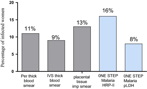
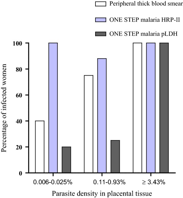
Results: The prevalence of malaria was 16%, 7.5%, 11.5%, 8% and 13% for One Step malaria HRP-II and pLDH RDTs, peripheral blood smear, IVS blood and placental tissue impression smears, respectively. The proportion of women positive by One Step malaria pLDH RDT and Pbs increased with parasite density in PTIS, while One Step malaria HRP-II RDT detected high proportion of infected women even with low parasite density. Although the prevalence of malaria infection by both microscopy and RDTs decreased significantly with mother age (0.0008 ≤ p ≤ 0.025), parity seemed to have very little influence. The sensitivity of One Step malaria HRP-II and pLDH RDTs were 96.15% and 61.53%, respectively, compared to 80.76% for Pbs (p = 0.014 and 0.0029, respectively). The specificity of these RDTs was 96.49% and 100%, respectively, compared to 100% for Pbs (p ≥ 0.12). In addition, the positive predictive values were 80.64% and 100% for HRP-II and pLDH-based RDTs, respectively, compared to 100% for Pbs (p < 0.0001 and 1, respectively), while the negative predictive values were 99.40% and 94.48%, respectively, compared to 97.16% for Pbs (p ≥ 0.49). The combination of One Step malaria HRP-II RDT and Pbs showed the similar performance as that observed with One Step malaria HRP-II RDT only.
Conclusion: These results depict One Step malaria HRP-II RDT to be better in detecting placental P. falciparum infection in pregnant women compared to Giemsa-stained peripheral thick blood smear. This is important for better case management since microscopic examination of PTIS cannot be employed during pregnancy.
Keywords: Cameroon; Peripheral blood; Placental tissue; Plasmodium falciparum; Pregnant women; RDT.
Figures


References
-
- World Malaria Report 2016. Geneva: World Health Organization; 2016.
-
- Salanti A, Staalsoe T, Lavstsen T, Jensen ATR, Sowa MPK, Arnot DE, et al. Selective upregulation of a single distinctly structured var gene in chondroitin sulphate A-adhering P. falciparum involved in pregnancy-associated malaria. Mol Microbiol. 2003;49:179–191. doi: 10.1046/j.1365-2958.2003.03570.x. - DOI - PubMed
-
- WHO . Guidelines for the treatment of malaria. Geneva: World Health Organization; 2010. - PubMed
MeSH terms
Substances
Grants and funding
LinkOut - more resources
Full Text Sources
Medical
