Review
. 2018 Dec 4;18(1):45.
doi: 10.1186/s40644-018-0180-6.

Endometrial cancer: an overview of novelties in treatment and related imaging keypoints for local staging

Affiliations
Review

Endometrial cancer: an overview of novelties in treatment and related imaging keypoints for local staging

Stefania Rizzo et al. Cancer Imaging. .

Abstract

Endometrial cancer is the most common gynaecologic malignancy in developed countries and its incidence is increasing. First-level treatment, if no contraindicated, is based on surgery. Pre-operative imaging is needed for evaluation of local extent and detection of distant metastases in order to guide treatment planning. Radiological evaluation, based on transvaginal ultrasound, MR and CT, can make the difference in disease management, paying special attention to assessment of entity of myometrial invasion, cervical stromal extension, and assessment of lymph nodal involvement and distant metastases.

Keywords: Cervical invasion; DWI; Endometrial cancer; MR; Myometrial invasion; TVUS.


Conflict of interest statement

Ethics approval and consent to participate

Not applicable.

Consent for publication

Not applicable.

Competing interests

The authors declare that they have no competing interests.

Publisher’s Note

Springer Nature remains neutral with regard to jurisdictional claims in published maps and institutional affiliations.

Figures

Fig. 1
Fig. 1
A 52-year-old woman with endometrioid adenocarcinoma (stage IA). Transvaginal ultrasound shows the endometrial cancer invading < 50% of the myometrium
Fig. 2
Fig. 2
A 51-year-old woman with endometrioid adenocarcinoma invading < 50% of the myometrium. Sagittal T2-w MRI (a) shows an intermediate-signal intensity tumour with disruption of the low signal junctional zone but the extent of myometrial invasion is unclear. Corresponding axial DWI image (b) shows an area of high signal intensity within the endometrial tumour invading only the inner myometrium (arrow)
Fig. 3
Fig. 3
A 66-year-old woman with endometrioid adenocarcinoma, grade 3. Sagittal T2-weighted image shows an intermediate-signal intensity endometrial tumour invading > 50% of the myometrium (a), with disruption of the low T2 signal junctional zone (white asterisk) and preservation of the low signal band of the outer myometrium (black arrowhead). Sagittal post contrast CT (b) in the same patient, performed for evaluation of distant metastases, does not discriminate the extent of myometrial infiltration
Fig. 4
Fig. 4
A 75-year-old woman with endometrioid carcinoma. Paracoronal post-gadoliunium T1-w subtracted dynamic images at 0 s (a), 60s (b) and 90s (c) help the evaluation of deep myometrial invasion in a post-menopausal patient
Fig. 5
Fig. 5
A 67-year-old patient with endometrioid carcinoma of the uterus, grade 2. Axial T2-weighted-DWI fused image shows an overlap of high DWI signal intensity and thicker uterine wall in the right side of the uterus. This makes the detection of tumour depth easier and more confident
Fig. 6
Fig. 6
Images of a 56-year-old woman with biopsy proved prostate cancer: multiparametric study: T2WI (a). FOCUS DWI images of b = 1000 s/mm2 (b and c). Perfusion map T2WI on sagittal plane (d) shows endometrial lesion confined to the endometrial cavity. FOCUS DWI images show tumor lesion in higher spatial resolution with a definite clear border, less artifacts, and image blurring
Fig. 7
Fig. 7
A 61-year-old women with endometrioid adenocarcinoma, grade 2. TVUS shows an exophytic isoechoic lesion extending to the cervix, as confirmed by post-surgical pathology
Fig. 8
Fig. 8
A 54-year-old women with EC. Sagittal (a) and axial (b) T2-w images show infiltration of the cervical stroma, without extension to the parametria, staging this tumour as II
Fig. 9
Fig. 9
A 67-year-old woman with EC stage IIIA. MR sagittal T2-w image depicting the endometrial lesion of the posterior uterine wall extending to the internal orifice without cervical involvement. There is a disruption of the junctional zone (arrowhead) indicating myometrial invasion, and disruption of the hypointense line contouring the myometrium on the outer side of the uterus (arrow), indicating serosal invasion. Collaterally there is a submucosal leiomyoma (asterisk)
Fig. 10
Fig. 10
A 54-year-old EC patient with deep myometrial infiltration and serosal involvement. Coronal (a) and sagittal (b) MR images show a small mass in the right ovary (arrows) with solid components of the same signal intensity as the EC, representing ovarian involvement
Fig. 11
Fig. 11
A 57-year-old woman with EC stage IIIB. Para-axial T2-w MR image (a) shows the endometrial carcinoma extending to the left parametrium (arrow). The tumour infiltrates the cervix through the internal uterine ostium leading to a retrodilation of the cavity which appears occupied by fluid (asterisk). In the same patient, the axial CT image shows the mass invading the left parametrium (b); collaterally there is a centimetric leiomyoma of the left wall of the uterus
Fig. 12
Fig. 12
A 46-year-old woman with EC grade 3. TVUS image (a) shows an endometrial mass of 4.79 cm (measure in yellow) invading < 50% of the myometrium. Despite a superficial myometrial invasion, CT images at staging (b) showed pelvic and lombo-aortic lymphoadenopathies (arrow), that bwere confirmed at surgery
Fig. 13
Fig. 13
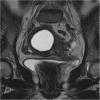
A 57-year-old woman with EC stage IVA. Coronal T2-weighted MR image showing a slightly hypointense tissue extending from the left parametrium to focally disrupt the urinary bladder wall on the left side (arrow)

References

    1. Siegel RL, Miller KD, Jemal A. Cancer statistics. CA Cancer J Clin. 2016;66(1):7–30. doi: 10.3322/caac.21332. - DOI - PubMed
    1. Colombo N, Creutzberg C, Amant F, et al. ESMO-ESGO-ESTRO endometrial consensus conference working group. ESMO-ESGO-ESTRO consensus conference on endometrial cancer: diagnosis, treatment and follow-up. Ann Oncol. 2016;27(1):16–41. doi: 10.1093/annonc/mdv484. - DOI - PubMed
    1. Pecorelli S. Revised FIGO staging for carcinoma of the vulva, cervix, and endometrium. Int J Gynaecol Obstet. 2009;105(2):103–104. doi: 10.1016/j.ijgo.2009.02.012. - DOI - PubMed
    1. Morice P, Leary A, Creutzberg C, Abu-Rustum N, Darai E. Endometrial cancer. Lancet. 2016;387(10023):1094–1108. doi: 10.1016/S0140-6736(15)00130-0. - DOI - PubMed
    1. Cragun JM, Havrilesky LJ, Calingaert B, et al. Retrospective analysis of selective lymphadenectomy in apparent early-stage endometrial cancer. J Clin Oncol. 2005;23(16):3668–3675. doi: 10.1200/JCO.2005.04.144. - DOI - PubMed