. 2018 Dec 4;9(6):e02204-18.
doi: 10.1128/mBio.02204-18.

Microbiota and Pathogen Proteases Modulate Type III Secretion Activity in Enterohemorrhagic Escherichia coli

Affiliations

Microbiota and Pathogen Proteases Modulate Type III Secretion Activity in Enterohemorrhagic Escherichia coli

Elizabeth A Cameron et al. mBio. .

Abstract

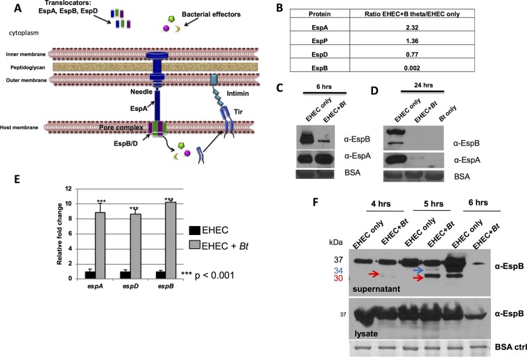
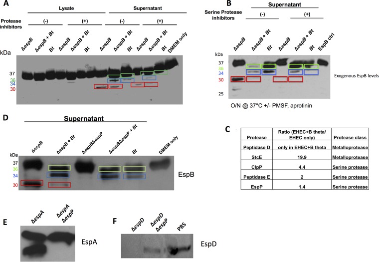
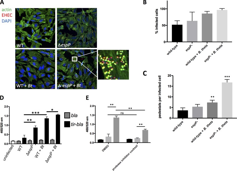
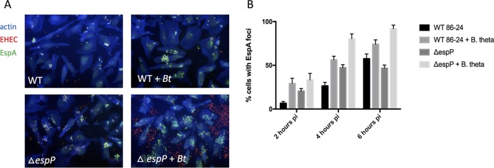
Enteric pathogens have complex interactions with the gut microbiota. Most of what is known about them has focused on microbiota-derived metabolites or small molecules that serve as nutrients and/or signals to aid in growth or transcriptionally regulate virulence gene expression. A common virulence strategy is to express a type III secretion system (T3SS), which is a molecular syringe deployed by many Gram-negative pathogens to hijack host cell function. Enterohemorrhagic Escherichiacoli (EHEC) requires its T3SS to colonize the intestinal tract and cause disease. Here we report that a prominent member of the intestinal microbiota, Bacteroides thetaiotamicron (Bt), secretes proteases that cleave the translocon of the T3SS of EHEC to enhance effector translocation into host cells. This is in contrast from an endogenous protease from EHEC itself (namely, EspP) that cleaves the translocon protein EspB in a different site to limit effector translocation. The EspB protein forms the T3SS pore in mammalian cells, and pore proteins are conserved in the T3SSs from several pathogens. This is the first demonstration of a commensal species directly processing a pathogen's T3SS, posing a new paradigm for how the microbiota can influence the severity of disease caused by bacterial pathogens. Because T3SSs are employed by many pathogens, this phenomenon has broad implications to commensal-pathogen relationships.IMPORTANCE The gut microbiota is usually regarded as providing colonization resistance against enteric pathogens. However, some pathogens evolved to thrive with the aid of certain members of the microbiota. Several Gram-negative bacteria employ type three secretion systems (T3SSs), which are molecular syringes that deliver effector proteins to host cells, hijacking host cell function. Here we show that the T3SS of enterohemorrhagic E. coli (EHEC) is cleaved by self and microbiota-derived proteases. Self-cleavage limits effector translocation, while cleavage by the microbiota member Bacteroides thetaiotamicron (Bt) exacerbates effector translocation and lesion formation on epithelial cells.

Keywords: Bacteroides; EspP; enterohemorrhagic E. coli (EHEC); microbiota; type three secretion.


Figures

FIG 1
FIG 1
The EHEC T3SS is cleaved by self-produced and Bacteroides thetaiotaomicron (Bt)-produced proteases. (A) EHEC T3SS: EspA is a sheath around the needle that connects to the pore proteins EspBD, forming the translocon. Several effectors, one of which is Tir, are translocated to the host cell. Tir serves as the receptor for the bacterial adhesin intimin and promotes actin polymerization, leading to AE lesion formation. (B) Mass spectrometric analysis of culture supernatants. Data are presented as a ratio of protein present in supernatants from EHEC grown in the presence of B. thetaiotaomicron (B theta) to protein present in supernatants from EHEC grown alone. (C and D) Western blot analysis of endogenous EspAB levels in culture supernatants of EHEC alone or EHEC plus Bt cultured for 6 h (C) and 24 h (D) in in vitro culture. EspAB levels in culture supernatants were analyzed by Western blotting using anti-EspAB antibodies. BSA is used as a loading control. α-EspB, anti-EspB antibody. (E) qPCR of espADB operon. espABD transcript levels from EHEC cultures with or without Bt were normalized to rpoA levels and are expressed as fold change over the values for EHEC alone. (F) Endogenous EspB levels. In vitro culture supernatants and lysates were collected at 4, 5, and 6 hours, and EspB was analyzed by Western blotting. The positions of the 37-kDa uncleaved EspB, 34-kDa cleaved EspB (blue arrows), and 30-kDa cleaved EspB (red arrows) are indicated. ctrl, control.
FIG 2
FIG 2
Both EHEC and Bt secrete proteases targeting EspB for degradation. (A) Western blot analysis of exogenous EspB. Purified rEspB-His was incubated with either the lysate or supernatant from EHEC ΔespB grown alone, EHEC ΔespB plus Bt, or Bt grown alone. Cultures were grown for 4.5 h prior to harvest. EspB-His (1 μg) was incubated at 37°C for 45 min in the presence or absence of a protease inhibitor cocktail (Sigma). (B) Exogenous EspB assay with serine protease inhibitors. rEspB-His (1 μg) was incubated with supernatants from specified cultures overnight in the presence or absence of serine protease inhibitors (PMSF, aprotinin) at 37°C. O/N, overnight. (C) Mass spectrometric analysis of proteases present in culture supernatant. Data are presented as a ratio of EHEC+Bt/EHEC alone. (D) Western blot analysis of exogenous EspB. Purified EspB-His was incubated with supernatants from EHEC ΔespB grown alone, EHEC ΔespB plus Bt, EHEC ΔespB ΔespP, EHEC ΔespB ΔespP plus Bt, or Bt grown alone. Cultures were grown for 4.5 h prior to harvest. EspB-His (1 μg) was incubated at 37°C for 45 min. DMEM medium was used as a negative control. (E) Western blot analysis of exogenous EspA. Purified EspA-His was incubated with supernatants from EHEC ΔespA or EHEC ΔespA ΔespP. (F) Western blot analysis of exogenous EspD. Purified EspD-His was incubated with supernatants from EHEC ΔespD, EHEC ΔespD ΔespP, or PBS as a negative control. Different EspB cleaved products are shown boxed in different colors: green, 36-kDa product; blue, 34-kDa product; and red, 30-kDa product.
FIG 3
FIG 3
EspB cleavage affects T3SS function. (A) Fluorescein actin staining (FAS) analysis. HeLa cells were infected with mCherry-expressing EHEC (WT or ΔespP) with or without Bt. At six hours postinfection, cells were washed, fixed, and stained with DAPI and FITC-phalloidin to visualize actin. Pedestals were visualized as green puncta localized with red bacteria. (B) Quantification of percent infected cells (percentage of cells with EHEC forming pedestals). **, P < 0.01; ***, P < 0.001. (C) Quantification of number of pedestals. The number of pedestals per infected cell was determined for each field, with each field containing approximately 20 cells. The averages and standard deviation across the fields (n = 4) are shown. **, P < 0.01; ***, P < 0.001. (D) Tir translocation assay. A TEM-1 β-lactamase system was used to measure translocation of Tir. Reporter EHEC strains express either TEM-1 β-lactamase (bla) as a control or a tir-bla fusion that will be translocated into host cells via the T3SS. HeLa cells were infected with reporter strains (WT or ΔespP) with or without Bt. Cells were then loaded with a fluorescent β-lactam compound whose emission spectrum was altered by β-lactamase cleavage. The 460/520 emission ratio reflects the level of β-lactamase activity and therefore the level of Tir translocation into cells. *, P < 0.05; **, P < 0.01; ***, P < 0.001. (E) The Tir translocation assay described for panel D was performed using a WT EHEC reporter strain +/− Bt in the presence of a protease inhibitor cocktail or DMSO vehicle control. **, P < 0.01; ns, not significant.
FIG 4
FIG 4
EspB proteolysis affects EspA filament formation. EspA immunofluorescence. HeLa cells were infected with mCherry-expressing EHEC (WT or ΔespP) with and without Bt. At 2, 4, and 6 h postinfection, cells were fixed, permeabilized, and stained for actin (blue) and EspA filaments (green). Representative images of the 6-h time point are shown (A) and the percentage of HeLa cells that contained EspA foci are quantified (B). The percentage of EspA-positive cells was quantified for 7 randomly selected fields containing 20 to 30 cells in each field, and the average and standard deviation are shown. The experiment was performed three times to ensure reproducibility. pi, postinoculation.

Comment in

References

    1. Baumler AJ, Sperandio V. 2016. Interactions between the microbiota and pathogenic bacteria in the gut. Nature 535:85–93. doi:10.1038/nature18849. - DOI - PMC - PubMed
    1. Scallan E, Hoekstra RM, Angulo FJ, Tauxe RV, Widdowson MA, Roy SL, Jones JL, Griffin PM. 2011. Foodborne illness acquired in the United States–major pathogens. Emerg Infect Dis 17:7–15. doi:10.3201/eid1701.091101p1. - DOI - PMC - PubMed
    1. Tuttle J, Gomez T, Doyle MP, Wells JG, Zhao T, Tauxe RV, Griffin PM. 1999. Lessons from a large outbreak of Escherichia coli O157:H7 infections: insights into the infectious dose and method of widespread contamination of hamburger patties. Epidemiol Infect 122:185–192. doi:10.1017/S0950268898001976. - DOI - PMC - PubMed
    1. Pacheco AR, Curtis MM, Ritchie JM, Munera D, Waldor MK, Moreira CG, Sperandio V. 2012. Fucose sensing regulates bacterial intestinal colonization. Nature 492:113–117. doi:10.1038/nature11623. - DOI - PMC - PubMed
    1. Curtis MM, Hu Z, Klimko C, Narayanan S, Deberardinis R, Sperandio V. 2014. The gut commensal Bacteroides thetaiotaomicron exacerbates enteric infection through modification of the metabolic landscape. Cell Host Microbe 16:759–769. doi:10.1016/j.chom.2014.11.005. - DOI - PMC - PubMed

Publication types

MeSH terms

LinkOut - more resources