Review
. 2019 Feb;18(2):139-158.
doi: 10.1038/s41573-018-0003-2.

Therapeutic strategies for sickle cell disease: towards a multi-agent approach

Affiliations
Review

Therapeutic strategies for sickle cell disease: towards a multi-agent approach

Marilyn J Telen et al. Nat Rev Drug Discov. 2019 Feb.

Abstract

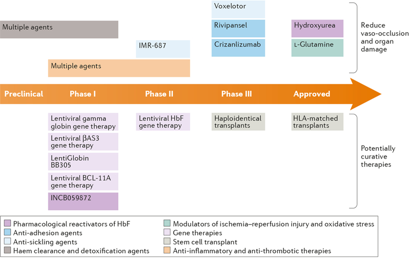
For over 100 years, clinicians and scientists have been unravelling the consequences of the A to T substitution in the β-globin gene that produces haemoglobin S, which leads to the systemic manifestations of sickle cell disease (SCD), including vaso-occlusion, anaemia, haemolysis, organ injury and pain. However, despite growing understanding of the mechanisms of haemoglobin S polymerization and its effects on red blood cells, only two therapies for SCD - hydroxyurea and L-glutamine - are approved by the US Food and Drug Administration. Moreover, these treatment options do not fully address the manifestations of SCD, which arise from a complex network of interdependent pathophysiological processes. In this article, we review efforts to develop new drugs targeting these processes, including agents that reactivate fetal haemoglobin, anti-sickling agents, anti-adhesion agents, modulators of ischaemia-reperfusion and oxidative stress, agents that counteract free haemoglobin and haem, anti-inflammatory agents, anti-thrombotic agents and anti-platelet agents. We also discuss gene therapy, which holds promise of a cure, although its widespread application is currently limited by technical challenges and the expense of treatment. We thus propose that developing systems-oriented multi-agent strategies on the basis of SCD pathophysiology is needed to improve the quality of life and survival of people with SCD.


Figures

Figure 1 |
Figure 1 |. Pathophysiology of sickle cell disease.
a | Sickle RBC oxidant and membrane changes. Upon deoxygenation of hemoglobin-S (Hb S), deoxygenated Hb S aggregates densely into polymers, and the red cell changes shape (“sickles”) due to this polymer-induced distortion. Spontaneous auto-oxidation of Hb S Fe2+ leads to the formation of Fe3+-methemoglobin and release of heme from the globin. O2 is reduced to superoxide and superoxide mutase (SOD) reacts to form H2O2. A catalytic cycle between ferric Hb-Fe3+ and the ferryl HbFe4+ can be initiated by H2O2, which is subsequently eliminated in a pseudoperoxidase-like manner. Ferryl Fe4+ Hb S promotes βCys93 oxidation, Hb dimerization and hemichrome formation. Upregulated NADPH-oxidase contributes to the oxidative environment, as do down-regulated oxidative defense pathways. Sickle red cells also have retained mitochondria, providing another source of oxidants that lead to membrane damage. There is a decrease in glutathione, as well as reduced activity of glutathione peroxidase, vitamin E, catalase, and peroxiredoxin. Oxidative membrane protein and lipid changes, including phosphatidyl serine (PS) expression on the membrane, promote erythrophagocytosis, adhesion, complement activation and prothombinase assembly. Excess oxidants lead band 3 sulfhydryl oxidation, enhanced erythrophagocytosis, altered Na+,Ca2+, and K+ homeostasis, dehydration, mechanosensitivity and deformability, increased fragility and vesiculation of microvesicles (that contain Hb S and heme), increased activity of adhesion receptors on the red blood cell membrane, and hemolysis.b | Hemolysis results in several intravascular events promoting vaso-occlusion in SCD. The abnormal sickle RBC membrane changes utimately lead to hemolysis within the vasculature and the release into the plasma of hemoglobin, reactive oxygen species (ROS) and arginase, which can accentuate nitric oxide (NO) depletion in the plasma. Free hemoglobin can react with NO and can readily oxidize to methemoglobin (MetHb); ROS can activate the endothelium of the vessel wall to promote adhesion molecule expression and the adherence of SRBCs, white cells (WBCs), and platelets. Within the vessel, free methemoglobin readily gives up its heme, which can interact with inflammatory cells and the endothelium. Excess plasma hemoglobin and heme depletes the heme scavengers haptoglobin and hemopexin. The adhesion of SRBCs, WBCs and platelets slow the flow in postcapillary venules, leading to further SRBC deoxygenation, sickling, and vaso-occlusion.
Figure 2 |
Figure 2 |. Sickle cell disease pathophysiological pathways and opportunities for targeted therapy.
The presence of sickle red blood cells (SRBCs) that contain hemoglobin S (HbS), along with SRBC-derived microparticles, are central to the pathophysiology of SCD and have a plethora of effects, ultimately leading to SRBC sickling, hemolysis, adhesive cell-cell interactions, inflammation, activation of coagulation, and endothelial dysfunction, as shown here. Cellular dehydration and deoxygenation along with hemoglobin S are at the center, leading to RBC sickling, hemolysis and release of free hemoglobin. Shown in the bottom left, free hemoglobin has several effects, including causing oxidant injury and endothelial activation. Abnormal membrane characteristics of SRBCs also activate coagulation processes that can then further activate endothelial cells, at bottom center, and inflammation, at bottom right. The SRBCs also adhere abnormally to leukocytes, other SRBCs, and the endothelial (shown on right). Leukocyte activate serves to proposgate the vaso-occlusive event, as well as causing ischemia/reperfusion injury. In each instance, investigators are working to develop agents to counteract these pathways (indicated in red).
Figure 3 |
Figure 3 |. The pipeline of sickle cell disease therapies.
The figure indicates promising therapeutic strategies and specific candidate therapeutics that are being actively pursued, as well as the two approved therapies (hydroxyurea and L-glutamine), classified as in the main text. Agents that are intended to amelioriate the various consequences of abnormal sickle red cells are shown in the upper half of the figure, and agents and approaches that coud potentially cure sickle cell disease are shown in the bottom half of the figure.
None

References

    1. Bunn HF Pathogenesis and treatment of sickle cell disease. The New England journal of medicine 337, 762–769, doi:10.1056/NEJM199709113371107 (1997). - DOI - PubMed
    1. Rees DC, Williams TN & Gladwin MT Sickle-cell disease. Lancet 376, 2018–2031, doi:10.1016/S0140-6736(10)61029-X (2010). - DOI - PubMed
    1. Platt OS et al. Mortality in sickle cell disease. Life expectancy and risk factors for early death [see comments]. New England Journal of Medicine 330, 1639–1644 (1994). - PubMed
    1. Kauf TL, Coates TD, Huazhi L, Mody-Patel N & Hartzema AG The cost of health care for children and adults with sickle cell disease. Am J Hematol 84, 323–327, doi:10.1002/ajh.21408 (2009). - DOI - PubMed
    1. Elmariah H et al. Factors associated with survival in a contemporary adult sickle cell disease cohort. American journal of hematology 89, 530–535, doi:10.1002/ajh.23683 (2014). - DOI - PMC - PubMed

Publication types

Substances