Review
. 2019 Jan;17(2):110-123.
doi: 10.1038/s41579-018-0117-x.

Viral RNA structure-based strategies to manipulate translation

Affiliations
Review

Viral RNA structure-based strategies to manipulate translation

Zane A Jaafar et al. Nat Rev Microbiol. 2019 Jan.

Abstract

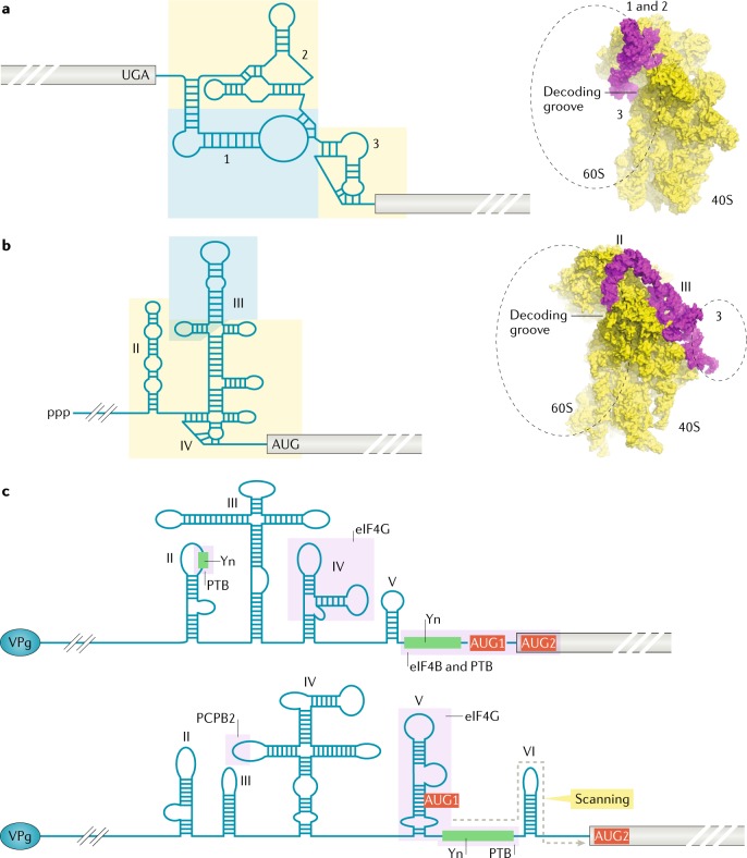
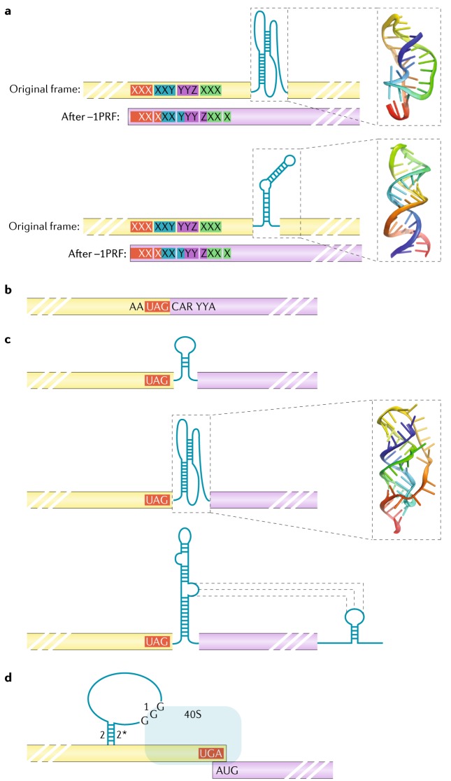
Viruses must co-opt the cellular translation machinery to produce progeny virions. Eukaryotic viruses have evolved a variety of ways to manipulate the cellular translation apparatus, in many cases using elegant RNA-centred strategies. Viral RNAs can alter or control every phase of protein synthesis and have diverse targets, mechanisms and structures. In addition, as cells attempt to limit infection by downregulating translation, some of these viral RNAs enable the virus to overcome this response or even take advantage of it to promote viral translation over cellular translation. In this Review, we present important examples of viral RNA-based strategies to exploit the cellular translation machinery. We describe what is understood of the structures and mechanisms of diverse viral RNA elements that alter or regulate translation, the advantages that are conferred to the virus and some of the major unknowns that provide motivation for further exploration.


Conflict of interest statement

The authors declare no competing interests.

Figures

Fig. 1
Fig. 1. Antiviral responses involving translation and viral RNA-based strategies to manipulate translation.
The translation cycle is generally divided into four phases, depicted here. For clarity, details such as each GTP hydrolysis event, all involved factors and individual steps are not shown; further details can be found in refs. Briefly, during canonical cap-dependent eukaryotic translation initiation, mRNA is recognized by the eukaryotic initiation factor (eIF) eIF4F complex, which contains eIF4E, eIF4G and eIF4A. This complex binds the modified nucleotide cap on the 5′ end of the mRNA, resulting in an mRNA activated for translation. A series of intermolecular recognition events leads to recruitment of the 43S complex to this activated mRNA; the 43S complex contains the small (40S) ribosomal subunit, eIFs (eIF3, eIF1, eIF1A and eIF5) and the eIF2–Met-tRNAiMet–GTP ternary complex. Next, the mRNA sequence is scanned in a 5′ to 3′ direction by the ribosomal subunit and associated factors in an ATP hydrolysis-dependent process. During scanning, eIF2-bound GTP is hydrolysed (stimulated by eIF5). The purpose of this scanning is to locate the proper start codon; the most used is an AUG triplet. When a start codon is selected, a codon–anticodon interaction is formed with Met-tRNAiMet in the P site, forming the 48S preinitiation complex. Phosphate is released by eIF2 and conformational changes involving a number of eIFs (eIF2, eIF1A, eIF1 and eIF5B) and a second GTP hydrolysis event on eIF5B lead to the release of most protein factors and the joining of the large (60S) ribosome subunit, creating an elongation-competent 80S ribosome. During elongation, codons are read by aminoacylated tRNAs delivered by the eukaryotic elongation factor (eEF) eEF1A in a GTP hydrolysis-dependent process. As tRNAs decode the message and enter the ribosome, they deliver their cognate amino acid to the growing polypeptide chain. Formation of each peptide bond is followed by GTP hydrolysis-dependent translocation by eEF2 and delivery of the next tRNA. Once a peptide chain has been made, the ribosome must terminate protein synthesis, release the protein and allow the ribosome to be used again (recycling). Once a stop codon (UAA, UGA or UAG) enters the A site, it is recognized by eukaryotic release factors (eRFs). The action of the eRFs along with other factors, including ATP-binding cassette sub-family E member 1 (ABCE1), ligatin and potentially others, leads to release of the peptide, subunit dissociation, tRNA release and ribosome recycling. During recycling, protein factors needed for the next round of translation are loaded back onto the ribosomal subunits; these include the proteins that make up the multifactor complex (MFC). Most phases of translation can be regulated, but two specific phases are noteworthy owing to their effect during viral infection, shown in purple boxes. The first is to interrupt the process of mRNA recruitment through the cap, primarily through the inactivation of eIF4E by hypophosphorylation of the factor or sequestration by eIF4E-binding proteins 1 and 2. The second is by inhibiting initiator tRNA delivery by phosphorylation of the α-subunit of eIF2. This prevents exchange of GDP for GTP on the factor; thus, it cannot be used to deliver initiator tRNA. Specific kinases do this in response to stresses induced by many viral infections, the most common being sensing of double-stranded RNA viral replication intermediates or endoplasmic reticulum stress by viral replication complexes. Viruses use RNA to interact with and exploit the translation process at many steps; examples that are discussed in this Review are shown in yellow boxes. CITE, cap-independent translation element; IRES, internal ribosome entry site.
Fig. 2
Fig. 2. Variations on scanning and start codon recognition.
Manipulation of the scanning and start codon recognition process by RNA sequence and structure is a strategy used by viruses to affect translation at the initiation phase and produce different proteins in different amounts. a | A canonical cellular message that is 7-methylguanosine-capped (cap) and has an AUG in a strong Kozak context is shown. Ribosomes scan (grey dashed line) until they reach this AUG, where translation of the ORF (pale yellow) begins, as shown with the thick black arrow. This mechanism results in efficient production of a single protein product (yellow, to the right). b | Leaky scanning can occur when multiple codons exist with different context strengths. In this conceptual diagram, ribosomes scan and then encounter a start codon in a weak context (shown here with pyrimidines (Y) at the −3 and +4 positions). Some ribosomes stop here and initiate, shown with the thin black arrow. Other ribosomes bypass this upstream AUG and continue scanning to reach a downstream AUG in a strong context and then initiate there. The amount of initiation at each AUG is dictated by the context strength. Two AUGs in the same reading frame are shown, leading to a longer and shorter isoform of the same protein, and the resultant population of protein products is shown to the right. c | Non-AUG codons can be used for initiation. An upstream alternative ORF is shown in blue, which is translated if scanning ribosomes initiate at a CUG in a weak context. Ribosomes that do not initiate at this CUG continue scanning to a downstream AUG in a strong context (pale yellow ORF). In the example shown here, the two ORFs overlap but are in different reading frames (green), leading to two entirely different protein products (right). Various combinations of overlapping reading frames in different contexts and start codons of different types and strengths can give rise to diverse outcomes in terms of types and relative amounts of protein, all from a single RNA template.
Fig. 3
Fig. 3. Internal ribosome entry sites.
a | Class 4 intergenic region internal ribosome entry sites (IRESs) are found between two viral ORFs. The three secondary structural domains are labelled. The yellow boxed area indicates the portion that interacts with the 40S subunit, and the blue boxed area is the portion that interacts with the 60S subunit. On the right is a cryo-electron microscopy (cryo-EM) reconstruction of the IRES bound to the 40S subunit (PDB 4V92), with the location of the domains labelled and the approximate location of the 60S subunit shown. The 60S subunit location is indicated, but that subunit has been removed to allow the domains of the IRES to be visualized. Domains 1 and 2 are labelled 1 and 2, as they form a single compact folded entity. b | Secondary structure cartoon of the hepatitis C virus IRES, representing the class 3 IRESs. The IRES is at the 5′ end of the viral genome, which starts with a triphosphate (ppp). Secondary structural domains are labelled, and the 40S and 60S subunit interaction sites are boxed in yellow and blue, respectively. At the right is a cryo-EM reconstruction of the IRES bound to the 40S subunit (PDB 5A2Q), with the location of the IRES RNA domains labelled and the approximate location of the 60S subunit shown; it has been removed to allow the full IRES to be seen. c | Class 1 and 2 IRESs are similar in organization and function but are not identical. Secondary structure cartoons of the encephalomyocarditis virus (class 2, top) and poliovirus (class 1, bottom) IRESs are shown, with secondary structure domains labelled. Both viral RNAs have a viral protein genome-linked (VPg) peptide on their 5′ end. Only the secondary structures necessary for IRES function are shown; upstream structures are omitted. The approximate binding sites for various eukaryotic initiation factors and IRES trans-acting factors are shown; additional details for related IRESs can be found in refs,. In the class 2 IRESs (top), there are two closely spaced start codons at the 3′ end of the IRES. For the class 1 IRESs (bottom), an upstream AUG codon (AUG1) is needed for ribosome entry, but then scanning leads to initiation at a downstream codon (AUG2). PCBP2, poly-C-binding protein 2; PTB, polypyrimidine tract-binding protein; Yn, polypyrimidine tract.
Fig. 4
Fig. 4. RNA elements within the coding region.
Specific folded RNA elements within a coding region can affect translation and lead to decoding of an alternative or extended frame. a | Programmed ribosomal frameshifting can be induced by structures that stall the ribosome and are coupled to slippery sequences. If encountered, RNA in the decoding groove can then shift into a different frame (in this example, a shift backwards of one nucleotide, in other words, a –1 programmed ribosomal frameshift (–1PRF)). If this occurs, translation continues but in a different frame (purple) than the original frame (yellow). Frameshifting can occur as a result of special pseudoknots (above) or stem-loops (below). NMR structures of a frameshifting pseudoknot from mouse mammary tumour virus (PDB 1RNK) or a hairpin from HIV-1 (PDB 1ZC5) are shown. b | Stop codon readthrough can occur through a variety of RNA-based mechanisms; when it occurs, the sequence downstream (purple) is translated. This simple case relies only on RNA sequence. Some tobamoviruses contain a stop codon within a UAG-CARYYA motif (R is a purine and Y is a pyrimidine) that can induce readthrough at low frequencies. c | Readthrough can be driven by structured RNA. A simple stem-loop (top). A pseudoknot from murine leukaemia gammaretrovirus and its NMR structure to the right (PDB 2LC8) (middle). The carnation Italian ringspot Tombusvirus readthrough signal involves long-range interactions (bottom). d | Termination upstream ribosome-binding sites (TURBSs) exist in some caliciviruses. They contain a stem (motifs 2 and 2*) with an extended loop (motif 1) that binds the ribosome on a terminating message to stimulate reinitiation on a downstream ORF (purple).
Fig. 5
Fig. 5. Translation enhancers in the 3′ UTR or at the 3′ end.
a | A class of 3′ cap-independent translation elements (3′-CITEs) called BTEs (barley yellow dwarf virus translation elements) adopt extended RNA architectures in the 3′ UTR, interacting with eukaryotic initiation factor 4G (eIF4G) and a stem-loop in the 5′ UTR to facilitate translation. b | PTEs (panicum mosaic virus-like translation enhancers) such as the one from saguaro cactus virus contain motifs that may pseudoknot (indicated by pk?) to form structures that recruit eIF4E and participate in long-range interactions with the sequence in either the 5′ UTR or the 5′ coding region. Other PTEs may interact with sequence in the 5′ coding region. c | I-shaped structures or Y-shaped structures (YSSs) recruit some component of the cap-binding complex and interact with sequences in the 5′ end of the mRNA (a YSS is shown). d | T-shaped structure (TSS)-type 3′-CITEs are proposed to bind directly to ribosomal subunits. e | TLSs (tRNA-like structures) are aminoacylated (AA, red), bound by eEF1A and/or interact with the ribosome. Crystal structure of the turnip yellow mosaic virus TLS is shown here (PDB 4P5J).

References

    1. Hinnebusch AG, Lorsch JR. The mechanism of eukaryotic translation initiation: new insights and challenges. Cold Spring Harb. Perspect. Biol. 2012;4:a011544. - PMC - PubMed
    1. Dever TE, Green R. The elongation, termination, and recycling phases of translation in eukaryotes. Cold Spring Harb. Perspect. Biol. 2012;4:a013706. - PMC - PubMed
    1. Nurenberg E, Tampe R. Tying up loose ends: ribosome recycling in eukaryotes and archaea. Trends Biochem. Sci. 2013;38:64–74. - PubMed
    1. Spriggs KA, Bushell M, Willis AE. Translational regulation of gene expression during conditions of cell stress. Mol. Cell. 2010;40:228–237. - PubMed
    1. Walsh D, Mathews MB, Mohr I. Tinkering with translation: protein synthesis in virus-infected cells. Cold Spring Harb. Perspect. Biol. 2013;5:a012351. - PMC - PubMed

Publication types