Clinical Trial
. 2018 Nov 20:9:2707.
doi: 10.3389/fimmu.2018.02707. eCollection 2018.

Lymphoid Stress Surveillance Response Contributes to Vitiligo Pathogenesis

Affiliations
Clinical Trial

Lymphoid Stress Surveillance Response Contributes to Vitiligo Pathogenesis

Liisi Raam et al. Front Immunol. .

Abstract

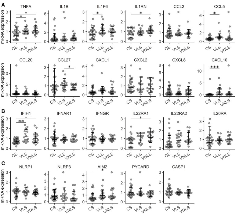
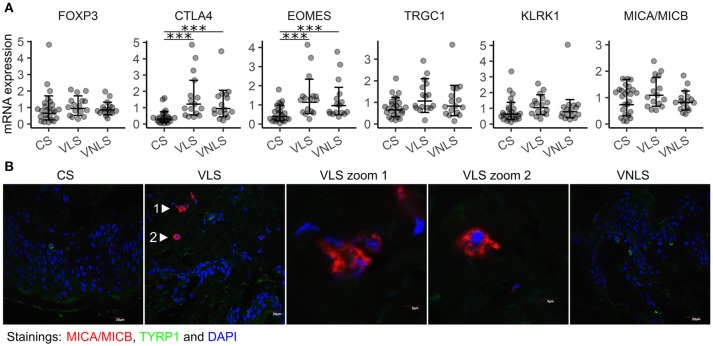
Vitiligo is a chronic multifactorial depigmentation disorder characterized by the destruction and functional loss of melanocytes. Although a direct cytotoxic T cell attack is thought to be responsible for melanocyte damage, the events leading to the loss of self-tolerance toward melanocytic antigens are not understood. This research aimed to identify novel cellular and molecular factors that participate in vitiligo pathogenesis through the application of gene expression and immunofluorescence analysis of skin biopsy samples along with immunophenotyping of circulating cells. Our study provides insights into the mechanisms involved in melanocyte destruction. The upregulation of stress-ligand MICA/MICB, recognized by activating receptors on innate and innate-like T cells, imply involvement of lymphoid stress surveillance responses in vitiligo lesions. A simultaneous increase in the expression of transcription factor EOMES that is characteristic for innate-like virtual memory T cells, suggest a similar scenario. Local lymphoid stress surveillance has been previously associated with the amplification of systemic humoral responses that were mirrored in our study by increased T follicular helper cells and switched memory B cell proportions in patients with active vitiligo. In addition, microtubule-associated protein light chain 3 staining was compatible with the activation of autophagy in keratinocytes and in the remaining melanocytes of vitiligo lesional skin.

Keywords: B cells; EOMES; LC3; MICA/MICB; WIPI1; autophagy; interferons; vitiligo.


Figures

Figure 1
Figure 1
Gene expression signature is consistent with a very mild inflammatory response in vitiligo. Relative expression of mRNAs encoding (A) cytokines, (B) receptors and (C) components of the inflammasome in the skin of control subjects (CS), and in the lesional skin (VLS) and non-lesional skin (VNLS) of vitiligo patients. Geometric mean ×÷ geometric standard deviation is indicated. *p < 0.05; **p < 0.01; and ***p < 0.001.
Figure 2
Figure 2
Concentration of cytokines in the plasma of control subjects (C) and vitiligo patients (V). Arithmetic mean ± standard deviation is indicated. *p < 0.05; **p < 0.01.
Figure 3
Figure 3
Innate-like cells and receptors are involved in vitiligo pathogenesis. (A) Relative mRNA expression of genes encoding markers of infiltrating immune cells and their target molecules. Geometric mean ×÷ geometric standard deviation is indicated. ***p < 0.001. (B) Immunofluorescence image of MICA/MICB (MHC class I chain-related protein A and B) and TYRP1 (tyrosinase related protein 1) in sections of biopsy samples from control subjects (CS), and in the lesional skin (VLS) and non-lesional skin (VNLS) of vitiligo patients. The images are representative of 3 healthy controls and 3 vitiligo patients.
Figure 4
Figure 4
Comparison of circulating lymphocyte subpopulations in vitiligo patients with different disease activity. (A) The percentage of regulatory T cells (Treg) among T helpers and unswitched memory (USM) B cells among B cells in vitiligo patients (V) and control individuals (C). (B) The percentage of naïve, switched memory (SM) and USM B cells among B cells and follicular helper T cells (Tfh) among T helper cells in vitiligo patients with active (A) Vs. stable (S) disease. Arithmetic mean ± standard deviation is indicated. *p < 0.05; **p < 0.01; and ***p < 0.001. (C) A scatter plot showing the correlation between the percentages of SM B cells and Tfh cells. Pearson's correlation coefficient (r) and the p-value of the association test are indicated on the plot.
Figure 5
Figure 5
WIPI1 expression and LC3 staining. (A) WIPI1 relative mRNA expression in the skin of control subjects (CS), and in the perilesional skin (VLS) and non-lesional skin (VNLS) of vitiligo patients. Geometric mean ×÷ geometric standard deviation is indicated. **p < 0.01; and ***p < 0.001. (B) WIPI1 relative mRNA expression in various cell types of the skin. Geometric mean ×÷ geometric standard deviation is indicated. 2D, two-dimensional; ALI, air-liquid interface; moLC, monocyte derived Langerhans cells. (C) Immunofluorescence image of LC3 (microtubule-associated protein 1A/1B-light chain 3) and TYRP1(tyrosinase related protein 1) in sections of biopsy samples from control subjects (CS), and lesional skin (VLS) and non-lesional skin (VNLS) of vitiligo patients. The white bar represents 5 μm. (D) Immunofluorescence image as in panel C with the white bar representing 20 μm. Neg. refers to a control slide with secondary antibodies only. (E) The box and whiskers plots show the median and interquartile range of the ratio between the fluorescence signal marking LC3 expression and DAPI (cell nuclei) in CS, VNLS, and VLS. Whiskers cover data points within a 1.5× interquartile range. *p < 0.05.

References

    1. Ezzedine K, Eleftheriadou V, Whitton M and van Geel N. Vitiligo. Lancet (2015) 386:74–84. 10.1016/s0140-6736(14)60763-7 - DOI - PubMed
    1. Silverberg JI, Silverberg NB. Association between vitiligo extent and distribution and quality-of-life impairment. JAMA Dermatol. (2013) 149:159–64. 10.1001/jamadermatol.2013.927 - DOI - PubMed
    1. Dahir AM, Thomsen SF. Comorbidities in vitiligo: comprehensive review. Int J Dermatol (2018) 57:1157-64. 10.1111/ijd.14055 - DOI - PubMed
    1. Boniface K, Seneschal J, Picardo M, Taieb A. Vitiligo: focus on clinical aspects, immunopathogenesis, and therapy. Clin Rev Allergy Immunol. (2018) 54:52–67. 10.1007/s12016-017-8622-7 - DOI - PubMed
    1. Jin Y, Andersen G, Yorgov D, Ferrara TM, Ben S, Brownson KM, et al. . Genome-wide association studies of autoimmune vitiligo identify 23 new risk loci and highlight key pathways and regulatory variants. Nat Genet. (2016) 48:1418–24. 10.1038/ng.3680 - DOI - PMC - PubMed

Publication types

MeSH terms