. 2018 Nov 20:9:1701.
doi: 10.3389/fpls.2018.01701. eCollection 2018.

Kinematical, Structural and Mechanical Adaptations to Desiccation in Poikilohydric Ramonda myconi (Gesneriaceae)

Affiliations

Kinematical, Structural and Mechanical Adaptations to Desiccation in Poikilohydric Ramonda myconi (Gesneriaceae)

Tim Kampowski et al. Front Plant Sci. .

Abstract

Resurrection plants have fascinated scientists since centuries as they can fully recover from cellular water contents below 10%, concomitantly showing remarkable leaf folding motions. While physiological adaptations have been meticulously investigated, the understanding of structural and mechanical adaptations of this phenomenon is scarce. Using imaging and bending techniques during dehydration-rehydration experiments, morphological, anatomical, and biomechanical properties of desiccation-tolerant Ramonda myconi are examined, and selected structural adaptations are compared to those of homoiohydrous Monophyllaea horsfieldii (both Gesneriaceae). At low water availability, intact and cut-off R. myconi leaves undergo considerable morphological alterations, which are fully and repeatedly reversible upon rehydration. Furthermore, their petioles show a triphasic mechanical behavior having a turgor-based structural stability at high (Phase 1), a flexible mechanically state at intermediate (Phase 2) and a material-based stability at low water contents (Phase 3). Lastly, manipulation experiments with cut-off plant parts revealed that both the shape alterations of individual structures, as well as, the general leaf kinematics largely rely on passive swelling and shrinking processes. Taken together, R. myconi possesses structural and mechanical adaptations to desiccation (in addition to physiological adaptations), which may mainly be passively driven by its water status influenced by the water fluctuations in its surroundings.

Keywords: desiccation tolerance; functional morphology; leaf folding kinematics; plant biomechanics; resurrection plants.


Figures

Figure 1
Figure 1
Habitus, leaf morphology and anatomy of R. myconi. (A) Cultivated flowering plants. (B) Adaxial and abaxial view of R. myconi leaves. Sampling areas for the location-specific RWC measurements are marked on the lamina and petiole of the right leaf. L1–L4: apical to basal lamina sampling areas [additional indices indicate locations left (1) or right (2) from the midrib]; P1–P4: apical to basal petiole sampling areas. (C) Cross-section of the petiole of R. myconi stained with TB. The image highlights all tissue types and the ROI used for the anatomical measurements in each thin section. ABE, abaxial epidermis; ADE, adaxial epidermis; LA, lacuna; LP, large-celled parenchyma (with pronounced lacunae); SP, small-celled parenchyma; TZ, transition zone between vascular and parenchymatous tissues; VC, vascular complex; VS, vascular strand. (D) Close-up of a longitudinal cut of the petiole showing the spiral thickenings of the xylem vessels (AO staining).
Figure 2
Figure 2
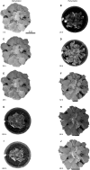
Water status dependent folding kinematics of R. myconi. Folding and shrinking of R. myconi plants at 0 h (A), after 24 h (C), 48 h (E), 225 h (G), and 450 h (I) of dehydration. Unfolding and swelling of R. myconi plants after 20 h (B), 28 h (D), 52 h (F), 68 h (H), and 80 h (J) of rehydration. R. myconi is able to withstand severe drought stress and re-establishes its normal shape upon rehydration. For reasons of simplification the graph depicts the results of one representative experiment.
Figure 3
Figure 3
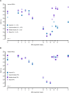
Location-specific RWC alterations of the lamina and petiole of R. myconi during DREs. (A) Different lamina sampling locations (circles) showed that the local RWCs during dehydration hardly differ. However, sometimes apical lamina parts recovered less than basal lamina parts during rehydration. (B) Local RWCs of petiole samples (squares) altered similarly throughout the complete DRE. For reasons of simplification the graph depicts the results of one representative experiment. The origin of each sample is color-coded: blue: apical sampling location; light-blue: apical-to-median sampling location; purple: median-to-basal sampling location; light-purple: basal sampling location. Further information on individual sampling locations is given in parentheses and Figure 1B. Dehydrated samples were collected from day 0 to day 12, whereas progressively rehydrated samples were gathered after last dehydration measurement on day 12.
Figure 4
Figure 4
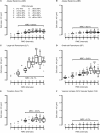
Tissue morphological alterations of R. myconi petioles during DREs. Cell area alterations of the adaxial epidermis (A), the abaxial epidermis (B), the large-celled parenchyma (C), the small-celled parenchyma (D), the transition zone between vascular and parenchymatous tissues (E), and the vascular system (F) in response to varying RWCs [100 to 0% RWC at 10% intervals, see legend in (A)]. The group sample sizes (below) and statistical references (above, Kruskal-Wallis test) are given for each boxplot. Outliers are presented as circles, single values as crosses. The median relative cell area changes (parentheses) are calculated from the cell areas at full turgescence (RWC interval 10) and those at the lowest RWC interval with N > 5 (usually interval 3, only for TZ interval 5 was used). ABE, abaxial epidermis; ADE, adaxial epidermis; LP, large-celled parenchyma; MRC, median relative cell area change; SP, small-celled parenchyma; TZ, transition zone between the vascular and the surrounding tissues; VC, vascular complex; VS, vascular system.
Figure 5
Figure 5
Water status dependent alterations of the petiolar biomechanics of R. myconi. Dehydration (open triangles) induces a triphasic mechanical behavior, which is characterized by a turgor-based structural stability at high and a material-based stability at low water contents, and is fully reversible upon rehydration (closed triangles). The structural bending elastic moduli (E) have been normalized to the median bending modulus at RWCs > 90%. The normalized bending elastic moduli (Enorm) of each RWC interval are either given as median ± standard error of the median (N ≥ 5, large dark-blue symbols), or as individual values (N < 5, small light-blue symbols).
Figure 6
Figure 6
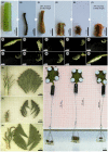
Assessment of structural adaptations to desiccation in R. myconi. (A–F) Comparative drying experiment using hypocotyl sections of desiccation-intolerant M. horsfieldii (A–C) and petiolar sections of desiccation-tolerant R. myconi (D–F) (both Gesneriaceae). Samples were imaged directly after sampling (A,D), after drying (B,E) and after rehydration (C,F). Hypocotyl and petiole segments regained 26 and 79% of their initial weights, respectively. (G–P) Image sequence illustrating the complex folding and shrinking behavior of a cut-off R. myconi leaf during dehydration (G–K), as well as, its recovery after submersion (L–P). The complex 3D folding-unfolding processes are structurally programmed into the leaf. (Q–S) Separated lamina and venation system parts, whose spatially complex folding motions were restricted to a 2D plane by clamping between two petri dishes, are shown in the hydrated state (Q), in the strongly contracted dehydrated state (R), and in the rehydrated state (S). The lamina is presumably responsible for (most of) the shrinkage of the leaf, whereas the first order veins are likely to channel the folding-unfolding movements. (T) During drying, the petioles exert contraction-induced forces high enough to pull weights.

References

    1. Alamillo J., Almoguera C., Bartels D., Jordano J. (1995). Constitutive expression of small heat shock proteins in vegetative tissues of the resurrection plant Craterostigma plantagineum. Plant Mol. Biol. 29, 1093–1099. 10.1007/BF00014981 - DOI - PubMed
    1. Aloni R. (1987). Differentiation of vascular tissues. Annu. Rev. Plant Physiol. Plant Mol. Biol. 38, 179–204. 10.1146/annurev.pp.38.060187.001143 - DOI
    1. Bartels D., Salamini F. (2001). Desiccation tolerance in the resurrection plant Craterostigma plantagineum. A contribution to the study of drought tolerance at the molecular level. Plant Physiol. 127, 1346–1353. 10.1104/pp.010765 - DOI - PMC - PubMed
    1. Baskin T. I. (2005). Anisotropic expansion of the plant cell wall. Annu. Rev. Cell Dev. Biol. 21, 203–222. 10.1146/annurev.cellbio.20.082503.103053 - DOI - PubMed
    1. Bateman R. M., Crane P. R., DiMichele W. A., Kenrick P. R., Rowe N. P., Speck T., et al. (1998). Early evolution of land plants: phylogeny, physiology, and ecology of the primary terrestrial radiation. Annu. Rev. Ecol. Evol. Syst. 29, 263–292. 10.1146/annurev.ecolsys.29.1.263 - DOI

LinkOut - more resources