. 2018 Nov 29:73:e562.
doi: 10.6061/clinics/2018/e562.

Proposal for a New Histological Scoring System for Cartilage Repair

Affiliations

Proposal for a New Histological Scoring System for Cartilage Repair

Maria Clara Ponce et al. Clinics (Sao Paulo). .

Abstract

Objective: This study aimed to develop a new histological scoring system for use in a partial-thickness cartilage repair animal model. Although previous papers have investigated the regeneration of articular cartilage, the good results achieved in small animals have not been replicated in large animal models or humans, possibly because of the frequent use of models with perforation of the subchondral bone plates. Partial-thickness lesions spare the subchondral bone, and this pattern is the most frequent in humans; therefore, new therapies should be tested using this model. However, no specific histological score exists to evaluate partial-thickness model results.

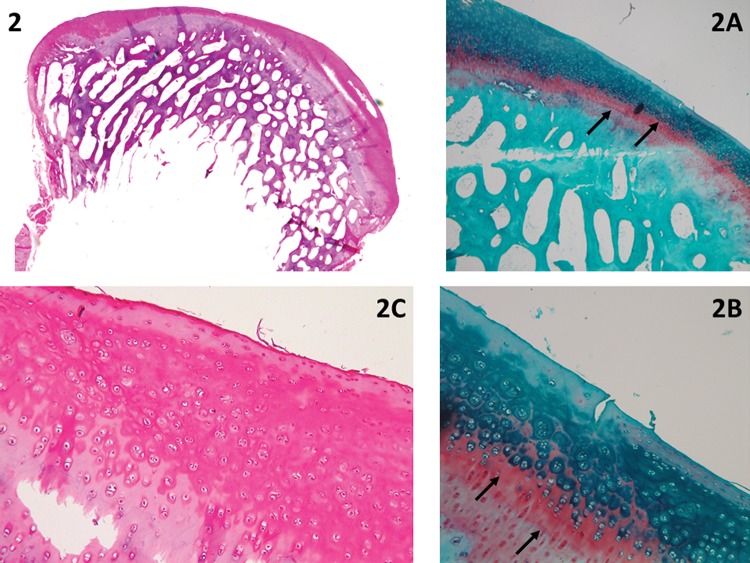
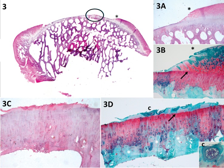
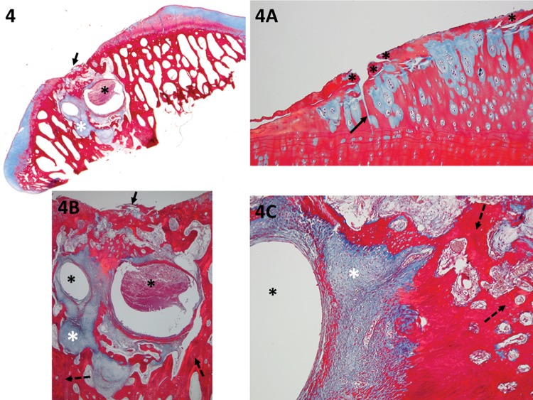
Methods: Histological sections from 30 ovine knees were reviewed to develop a new scoring system. The sections were subjected to H&E, Safranin O, and Masson's trichrome staining.

Results: This paper describes a new scoring tool that is divided into sections in detail: repair of tissue inside the lesion, cartilage around the lesion and degenerative changes at the base of the lesion. Scores range from 0 to 21; a higher score indicates better cartilage repair.

Discussion: Unlike existing tools, this new scale does not assign points for the positioning of a tidemark; we propose evaluation of the degenerative changes to the subchondral bone and calcified cartilage layer. It is necessary to remove the whole joint to access and study the evolution of the lesion as well as the surrounding tissue.

Conclusion: This article emphasizes the importance of a partial-thickness animal model of cartilage repair and presents a new histological scoring system.


Conflict of interest statement

No potential conflict of interest was reported.

Figures

Figure 1
Figure 1
A panoramic view (1A, H&E) of a piece of cartilage that was removed from the sample (black asterisk), shown in detail with Safranin O stain (1C). With the Masson's trichrome stain (1B), it is possible to distinguish between old (red) and new (blue) collagen; therefore, it can be assumed that this piece of cartilage (stained in blue) resulted from cartilage growth. Nevertheless, the score cannot be applied because a discreet detachment occurred. Furthermore, in 1A-1C, it is possible to observe small cartilage growth (black arrows), which can be evaluated with the UPT score; the horizontal growth occupies less than 20% of the defect (1 point), and the vertical growth (at its highest point) corresponds to less than 20% of the border height (1 point). Panel 1D (Safranin O) shows an example of cartilage hypocellularity with >25% of clusters and few isolated chondrocytes. Most of the cellularity is formed by clusters (white asterisk). The white arrows indicate the line formed between the pre-existing cartilage and the new cartilage (e.g., a “cementing line”), called a “tidemark.” Panel 1E (Safranin O) shows an example of relatively normal cellularity, in which the new cartilage follows the pre-existing tissue pattern of chondrocyte distribution.
Figure 2
Figure 2
The complete regeneration of the cartilage is shown in a panoramic view. The difference between the new and old cartilage is clear due to the evident line that separates the section, similar to a “tidemark” separating the pre-existing and new tissue, as shown by Safranin O staining (arrows in 2A and 2B). The cellularity in the new cartilage has a distribution pattern similar to that of the old cartilage (2C). The following evaluation was made: horizontal filling completed 100% of the defect (4 points); vertical filling at its highest point was equal to the border height (100%, equal to 4 points); <25% of chondrocytes formed clusters (cellularity=1 point); Safranin staining was heterogeneous (1 point); the new tissue was integrated into both borders (2 points); and residual cartilage both at the base and the subchondral bone was intact and normal (3 and 4 points, respectively); total=19 points.
Figure 3
Figure 3
The panoramic view shows a small amount of growth of new cartilage in the middle of the defect (circled area). Panel 3A* shows new cartilage growth at the lateral internal border, as confirmed by Safranin O staining in 3B* (heterogeneous and with a tidemark – arrow). Panels 3C and 3D (circled area in 3) show little cartilage growth in the middle of the lesion. According to the evaluation, horizontal filling accounted for less than 20% of the defect (1 point); vertical filling at its highest point was less than 20% of the border height (1 point); the hypocellularity was mild with <25% of clusters (evident in 3B and 3D-c, 1 point); Safranin O staining was heterogeneous (1 point); the new tissue was integrated into only one border (1 point – 3*, 3A* and 3B*); and the residual cartilage and subchondral bone (white *) were normal (3 and 4 points, respectively), total=12 points.
Figure 4
Figure 4
The panoramic view shows the exposition of the subchondral bone, likely due to an inadequate technique; therefore, the deep layers were not spared. The residual cartilage has fibrillation (black asterisk) and fissures (black arrow) (detail in 4A). Focal erosion is present (black arrow in 4 and 4B) (detail in 4B) (the lowest score should be considered in this case; thus, because of the presence of erosion, 1 point was assigned for the cartilage at the base of the lesion). The subchondral bone has a severe cystic lesion (black asterisk in 4, 4B and 4C) surrounded by loose, immature, fibrous connective tissue and new vessels (granulation tissue – white asterisk in 4, 4B and 4C). Because new sclerotic bone (detailed in 4B – dashed arrow) and remodeling bone formation (detailed 4C – dashed arrow) are observed, the total score for the subchondral bone is 1 point.

References

    1. Poole CA. Articular cartilage chondrons: form, function and failure. J Anat. 1997;191((Pt 1)):1–13. doi: 10.1046/j.1469-7580.1997.19110001.x. - DOI - PMC - PubMed
    1. Zorzi AR, Amstalden EM, Plepis AM, Martins VC, Ferretti M, Antonioli E, et al. Effect of human adipose tissue mesenchymal stem cells on the regeneration of ovine articular cartilage. Int J Mol Sci. 2015;16((11)):26813–31. doi: 10.3390/ijms161125989. - DOI - PMC - PubMed
    1. Knutsen G, Engebretsen L, Ludvigsen TC, Drogset JO, Grontvedt T, Solheim E, et al. Autologous chondrocyte implantation compared with microfracture in the knee. A randomized trial. J Bone Joint Surg Am. 2004;86-A((3)):455–64. doi: 10.2106/00004623-200403000-00001. - DOI - PubMed
    1. Saris DB, Vanlauwe J, Victor J, Haspl M, Bohnsack M, Fortems Y, et al. Characterized chondrocyte implantation results in better structural repair when treating symptomatic cartilage defects of the knee in a randomized controlled trial versus microfracture. Am J Sports Med. 2008;36((2)):235–46. doi: 10.1177/0363546507311095. - DOI - PubMed
    1. Verdonk P, Dhollander A, Almqvist KF, Verdonk R, Victor J. Treatment of osteochondral lesions in the knee using a cell-free scaffold. Bone Joint J. 2015;97-B((3)):318–23. doi: 10.1302/0301-620X.97B3.34555. - DOI - PubMed

LinkOut - more resources