

Case Reports
doi: 10.5935/abc.20180231.
Case 6 - Woman with Ischemic Heart Disease Admitted due to Chest Pain and Shock
[Article in
English,
Portuguese]
Affiliations
- PMID: 30517382
- PMCID: PMC6263468
- DOI: 10.5935/abc.20180231
Item in Clipboard
Case Reports
Case 6 - Woman with Ischemic Heart Disease Admitted due to Chest Pain and Shock
[Article in
English,
Portuguese]
Arq Bras Cardiol.
2018 Dec.
No abstract available
Figures

Electrocardiogram - Sinus rhythm, low voltage of the QRS complex in the
frontal plane, electrically inactive area in the inferior wall and left
bundle branch block.
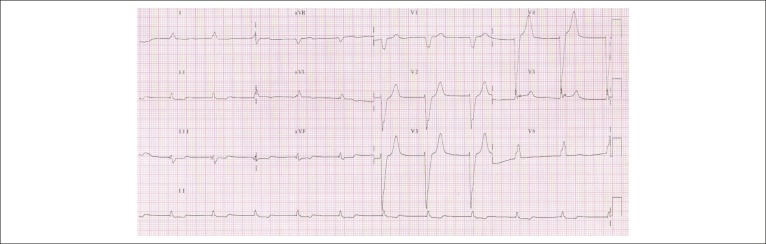
Electrocardiogram - Sinus rhythm, left bundle branch block and positive T
waves on an also positive derivative of the QRS complex.

Cross-sections of the heart at the level of the ventricles (short axis)
showing previous transmural infarctions in the anterior and septal walls
(arrows). These same places show thinning of the wall and, localized
slight dilatation (aneurysm). There is also a cavitary thrombus in the
ventricular apex (asterisk).

Right lung cross-section at its long axis showing the presence of
thromboembolism in the central branch of the pulmonary artery (arrow).
At the base, there are two triangular areas (asterisks) where the
parenchyma is homogeneous and reddish in color, corresponding to recent
pulmonary infarctions.

Photomicrography of the right pleura showing neutrophilic exudate on the
surface (asterisk), characterizing acute pleuritis. Hematoxylin-eosin
staining, objective magnification = 10X.
References
-
- Sgarbossa EB, Pinski SL, Barbagelata A, Underwood DA, Gates KB, Topol EJ. Electrocardiographic diagnosis of evolving acute myocardial infarction in the presence of left bundle-branch block GUSTO-1 (Global Utilization of Streptokinase and Tissue Plasminogen Activator for Occluded Coronary Arteries) Investigators. N Engl J Med. 1996;334(6):481–487. - PubMed
-
- Smith SW, Dodd KW, Henry TD, Dvorak DM, Pearce LA. Diagnosis of ST-elevation myocardial infarction in the presence of left bundle branch block with the ST-elevation to S-wave ratio in a modified Sgarbossa rule. Ann Emerg Med. 2012;60(6):766–776. - PubMed
-
- Kopterides P, Lignos M, Papanikolaou S, Papadomichalakis E, Mentzelopoulos S, Armaganidis A. Pleural effusion causing cardiac tamponade report of two cases and review of the literature. Heart Lung. 2006;35(1):66–67. - PubMed
Publication types
MeSH terms
LinkOut - more resources
Full Text Sources
Medical
