. 2018 Dec 4;7(12):180.
doi: 10.3390/antiox7120180.

Regulation of Oxidative Stress in Corneal Endothelial Cells by Prdx6

Affiliations

Regulation of Oxidative Stress in Corneal Endothelial Cells by Prdx6

Matthew Lovatt et al. Antioxidants (Basel). .

Abstract

The inner layer of the cornea, the corneal endothelium, is post-mitotic and unable to regenerate if damaged. The corneal endothelium is one of the most transplanted tissues in the body. Fuchs' endothelial corneal dystrophy (FECD) is the leading indication for corneal endothelial transplantation. FECD is thought to be an age-dependent disorder, with a major component related to oxidative stress. Prdx6 is an antioxidant with particular affinity for repairing peroxidised cell membranes. To address the role of Prdx6 in corneal endothelial cells, we used a combination of biochemical and functional studies. Our data reveal that Prdx6 is expressed at unusually high levels at the plasma membrane of corneal endothelial cells. RNAi-mediated knockdown of Prdx6 revealed a role for Prdx6 in lipid peroxidation. Furthermore, following induction of oxidative stress with menadione, Prdx6-deficient cells had defective mitochondrial membrane potential and were more sensitive to cell death. These data reveal that Prdx6 is compartmentalised in corneal endothelial cells and has multiple functions to preserve cellular integrity.

Keywords: Fuchs’ endothelial corneal dystrophy; Prdx6; cornea; lipid peroxidation; mitochondrial membrane potential.


Conflict of interest statement

The authors declare that they have no competing interests. The funders had no role in study design, data collection and analysis, decision to publish, or preparation of the manuscript.

Figures

Figure 1
Figure 1
(A,B) Prdx6 is predominantly expressed at the cell surface in human corneal endothelial cells (CEnCs). The indicated cells had their cell surface biotinylated using Sulfo-NHS-SS-Biotin. Cell lysates were subjected to immunoprecipitation with NeutrAvidin™ agarose. Eluted proteins were probed with Prdx6 antibodies. For comparative purposes, an aliquot of cell lysate prior to immunoprecipitation was used for analysis of total cellular Prdx6 levels. Bands were quantitated, and relative surface Prdx6 levels were normalised against total Prdx6 levels. Primary human corneal endothelial cell (pCEnC) levels were arbitrarily set to 100. Fib: corneal fibroblasts. RMECs: retinal microvascular endothelial cells.
Figure 2
Figure 2
Prdx6 levels are sensitive to oxidative stress. (A) Primary cultures of human CEnCs) were treated with tert-butyl hydroperoxide (tBHP) for 3 h. pCEnCs were biotinylated and cell surface proteins purified by immunoprecipitation. The flow-through, unbound fraction served as a control for membrane fractionation. (B) pCEnCs and (C) A549 cells were treated with tBHP for 3 h and plasma membrane was isolated by density dependent centrifugation. Data from three independent experiments ± SEM is shown. Prdx6 levels were normalized to Na+/K+-ATPase and expressed relative to untreated (−) controls. Student t-test was performed to evaluate statistical significance (** p-value < 0.005).
Figure 3
Figure 3
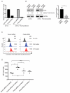
Targeted knockdown of Prdx6 in CEnCs reveals a role for Prdx6 in regulating lipid peroxidation. (A) Prdx6 mRNA levels were determined by qPCR analysis. Expression levels are shown relative to control siRNA treated B4G12 CEnCs and normalized to GAPDH. (B) Representative western blot analysis of aliquots of siRNA-transfected B4G12 CEnCs probed with anti-Prdx6 antibodies. Protein levels were quantitated relative to GAPDH and normalised to control siRNA. Data also expressed as mean ± SEM (n = 6). * p-value (<0.05) was determined by student t-test. (C) Lipid peroxidation was measured in B4G12 CEnCs incubated with linoleamide alkyne (LAA) in the presence (cumene hydroperoxide (CH) treated) or absence of CH (not treated). B4G12 CEnCs incubated in the absence of LAA served as negative controls. (D) Data are expressed as mean fluorescence intensity (MFI) from three independent experiments. Two-way ANOVA with multiple comparisons was used to test statistical significance. Asterix (*) indicates statistical significance; * p < 0.05, ** p < 0.005, *** p < 0.01, n.s.: no significant difference.
Figure 4
Figure 4
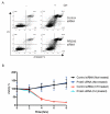
Normal apoptosis in the absence of Prdx6. (A) B4G12 CEnCs were treated with CH (100 μm) for 4 h and stained with Annexin V and propidium iodide (PI). Representative FACS plots are shown and the percentages of viable (AnVPI), early, (AnV+PI), late apoptotic (AnV+PI+), and necrotic (AnVPI+) are shown. (B) B4G12 CEnC cells were seeded on xCELLigence plates and continuously monitored for 8 h after the addition of CH (50 μm).
Figure 5
Figure 5
Prdx6-deficient cells are susceptible to cell death induced by menadione MN. (A) Representative FACS plots of TMRE mitochondrial membrane potential (ΔΨm) in siRNA-transfected B4G12 cells following treatment with 50 μm MN (90 min) or 50 μm CCCP (15 min). Percentage of cells with loss of ΔΨm is shown as well as overall TMRE mean fluorescence intensity (MFI). (B) Data are presented as mean values ± SEM (n = 4) of percentage TMRE negative. Two-way ANOVA reveals statistical significance (*p < 0.005) between MN treated control and Prdx6 siRNA-transfected cells. (C) Viability in response to MN and tBHP was assessed as in Figure 4B. Data is presented as mean values ± SEM (n = 3). Statistical analysis for each time point was carried out using two-way ANOVA with Bonferroni post-hoc multiple comparisons test (* p < 0.0001, ** p < 0.001).

References

    1. Peh G.S.L., Beuerman R.W., Colman A., Tan D.T., Mehta J.S. Human corneal endothelial cell expansion for corneal endothelium transplantation: An overview. Transplantation. 2011;91:811–819. doi: 10.1097/TP.0b013e3182111f01. - DOI - PubMed
    1. Wahlig S., Lovatt M., Mehta J.S. Functional role of peroxiredoxin 6 in the eye. Free Radic. Biol. Med. 2018;126:210–220. doi: 10.1016/j.freeradbiomed.2018.08.017. - DOI - PubMed
    1. Bourne W.M. Biology of the corneal endothelium in health and disease. Eye. 2003;17:912–918. doi: 10.1038/sj.eye.6700559. - DOI - PubMed
    1. Laing R.A., Sanstrom M.M., Berrospi A.R., Leibowitz H.M. Changes in the corneal endothelium as a function of age. Exp. Eye Res. 1976;22:587–594. doi: 10.1016/0014-4835(76)90003-8. - DOI - PubMed
    1. Galgauskas S., Norvydaitė D., Krasauskaitė D., Stech S., Ašoklis R.S. Age-related changes in corneal thickness and endothelial characteristics. Clin. Interv. Aging. 2013;8:1445–1450. doi: 10.2147/CIA.S51693. - DOI - PMC - PubMed

LinkOut - more resources