. 2018 Dec 4;19(12):3876.
doi: 10.3390/ijms19123876.

Fluorescently Tagged CCL19 and CCL21 to Monitor CCR7 and ACKR4 Functions

Affiliations

Fluorescently Tagged CCL19 and CCL21 to Monitor CCR7 and ACKR4 Functions

Vladimir Purvanov et al. Int J Mol Sci. .

Abstract

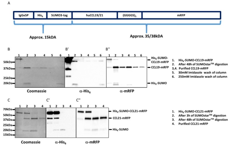
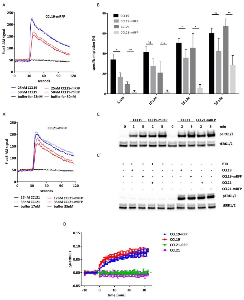
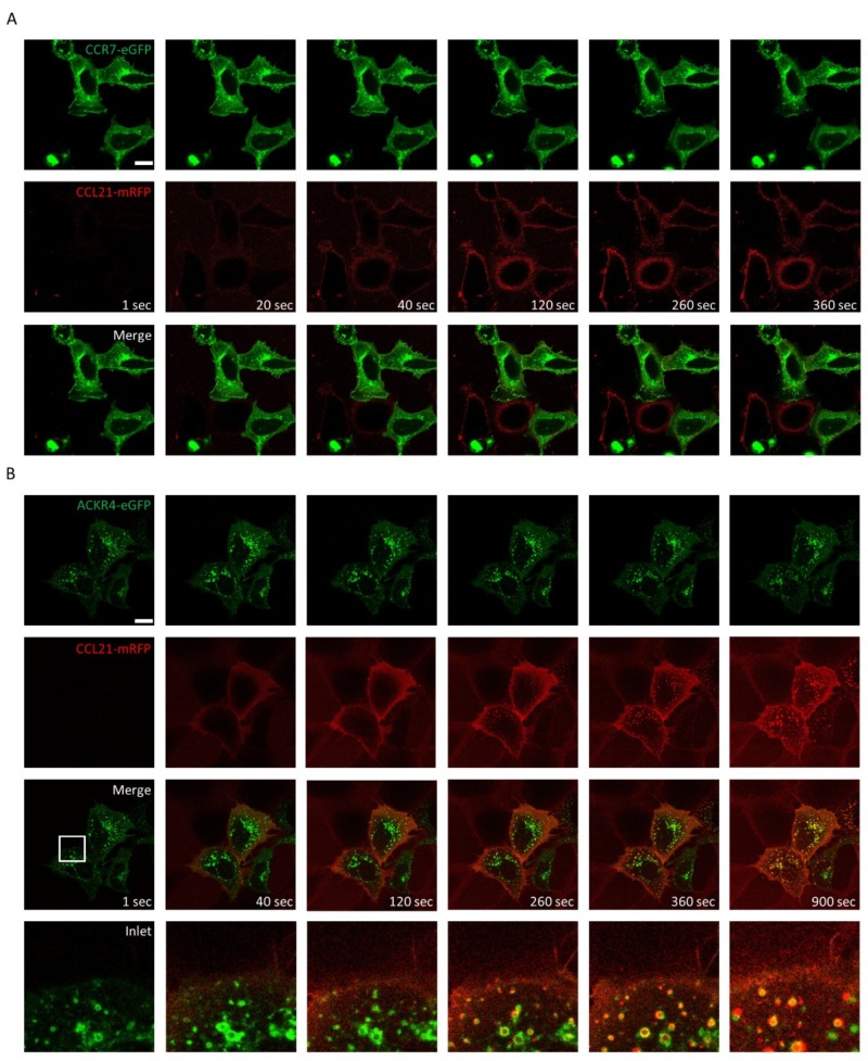
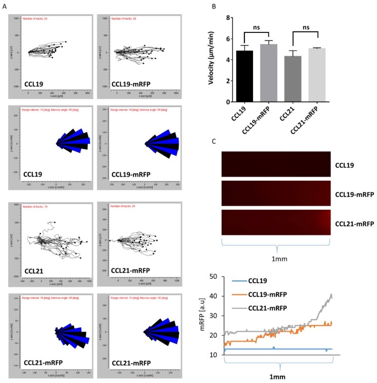
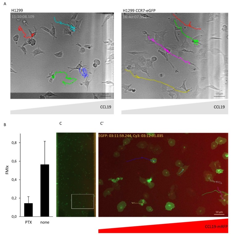
Chemokines are essential guidance cues orchestrating cell migration in health and disease. Cognate chemokine receptors sense chemokine gradients over short distances to coordinate directional cell locomotion. The chemokines CCL19 and CCL21 are essential for recruiting CCR7-expressing dendritic cells bearing pathogen-derived antigens and lymphocytes to lymph nodes, where the two cell types meet to launch an adaptive immune response against the invading pathogen. CCR7-expressing cancer cells are also recruited by CCL19 and CCL21 to metastasize in lymphoid organs. In contrast, atypical chemokine receptors (ACKRs) do not transmit signals required for cell locomotion but scavenge chemokines. ACKR4 is crucial for internalizing and degrading CCL19 and CCL21 to establish local gradients, which are sensed by CCR7-expressing cells. Here, we describe the production of fluorescently tagged chemokines by fusing CCL19 and CCL21 to monomeric red fluorescent protein (mRFP). We show that purified CCL19-mRFP and CCL21-mRFP are versatile and powerful tools to study CCR7 and ACKR4 functions, such as receptor trafficking and chemokine scavenging, in a spatiotemporal fashion. We demonstrate that fluorescently tagged CCL19 and CCL21 permit the visualization and quantification of chemokine gradients in real time, while CCR7-expressing leukocytes and cancer cells sense the guidance cues and migrate along the chemokine gradients.

Keywords: ACKR4; CCL19; CCL21; CCR7; cancer metastasis; cell migration; chemokine receptors; fluorescent chemokines; leukocytes.


Conflict of interest statement

The authors declare no conflict of interest. The funders had no role in the design of the study; in the collection, analyses, or interpretation of data; in the writing of the manuscript, or in the decision to publish the results.

Figures

Figure 1
Figure 1
Design and purification of CCL19-mRFP and CCL21-mRFP. (A) Graphic scheme of the constructs used to generate fluorescently tagged full-length chemokines. (B,C) Coomassie-stained SDS-PAGE gels (left) and corresponding Western blots (middle and right) of samples collected during the purification steps of CCL19-mRFP (B) and CCL21-mRFP (C). Secreted His6-SUMO-CCL19-mRFP was collected from the supernatants of transfected HEK293 cells and affinity purified over a Ni2+ column (B, line 1). The His6-SUMO tag was cleaved off by incubation for 48 h with SUMOstar™ protease at 8 °C (B, lane 2) and applied again on the Ni2+ column to remove the His6-SUMO tag. The purified CCL19-mRFP was collected as flow-through (B, lanes 3,4). The column was washed with 30mM imidazole (B, lane 5) and 250 mM imidazole to completely remove the His6-SUMO tag from the column for reuse (B, lane 6). Similarly, secreted His6-SUMO-CCL21-mRFP (C, lane 1), was digested with SUMOstar™ protease for 3 and 48 h (C, lanes 2,3) and applied to a Ni2+ column. Purified CCL21-mRFP was collected as flow-through (C, line 4). Western blots of the same samples using anti-His tag (B′,C′) or anti mRFP (B″,C″) antibodies are shown.
Figure 2
Figure 2
CCL19-mRFP and CCL21-mRFP are functional chemokines. (A) Real-time changes in [Ca2+]i concentrations in CCR7 transfected 300-19 cells in response to graded concentrations of CCL19 and CCL19-mRFP (A) and CCL21 and CCL21-mRFP (A′). A representative experiment out of three is shown. (B) Transwell chemotaxis of human mature MoDCs in response to chemokines. Human mature MoDCs were allowed to migrate in response to gradient concentrations of chemokines for 180 min. Migrated cells were counted and percentages of specifically migrated cells relative to the input were calculated. Mean values and SEM of duplicates derived from three independent experiments are shown. (C) Stimulation of human mature MoDCs with CCL19-mRFP and CCL21-mRFP results in the phosphorylation of ERK1/2. Human mature MoDCs were stimulated with 50 nM of CCL19, CCL19-mRFP or CCL21-mRFP for the indicated time points. ns: statistically not significant; * p < 0.05; ** p < 0.005. (C’) MoDCs were pre-treated with 200 ng/mL PTx for 3 h and subsequently stimulated with 50 nM of chemokines for 5 min. Phosphorylation of ERK1/2 (pERK1/2) was determined by Western blotting. Re-probing the blots for total ERK1/2 (tERK1/2) served as a control for equal protein loading. (D) Chemokine-induced β-arrestin recruitment to CCR7. β-arrestin recruitment was determined by BRET upon stimulation with 50 nM of indicated chemokine. Mean values ±S.D. of 2 (CCL21, CCL21-mRFP) or 3 (CCL19, CCL19-mRFP) independent experiments with technical duplicates are shown.
Figure 3
Figure 3
Internalization of CCL19-mRFP by CCR7-eGFP and ACKR4-eGFP. 100 nM of CCL19-mRFP was added to HEK293 cells transiently transfected with CCR7-eGFP (A) or ACKR4-eGFP (B) and CCL19-mRFP internalization was recorded by time-lapse video microscopy. Series of images captured at indicated time points are illustrated (scale bar: 10 µm). Frames: magnification of images.
Figure 4
Figure 4
Monitoring CCL21-mRFP binding to CCR7-eGFP and internalization by ACKR4-eGFP. 100nM of CCL21-mRFP was added to HEK293 cells transiently transfected with CCR7-eGFP (A) or ACKR4-eGFP (B) and CCL21-mRFP binding and receptor trafficking was recorded by time-lapse video microscopy. Series of images captured at indicated time points are depicted (scale bar: 10 µm). White frames: magnification of images.
Figure 5
Figure 5
DC migration in 3D collagen along gradients of CCL19-mRFP and CCL21-mRFP. Mouse mature bone-marrow-derived DCs were imbedded in 3D collagen in µ-slide chemotaxis chambers. CCL19, CCL19-mRFP, CCL21, or CCL21-mRFP at 100 nM concentration was applied to the right reservoir. (A) Combined rose diagrams and tracks of single cells migrating towards higher concentrations of chemokines from a representative experiment are shown. (B) Mean velocity of individually migrating cells derived from three independent experiments were quantified. ns: statistically not significant. (C) Gradients of CCL19-mRFP and CCL21-mRFP from (A) were visualized by fluorescence microscopy and enumerated by determining the fluorescent intensity derived from the corresponding mRFP signal.
Figure 6
Figure 6
CCR7-dependet migration of H1299 cancer cells along CCL19 and CCL19-mRFP gradients. (A) H1299 wild-type cells and H1299 cells expressing CCR7-eGFP were imbedded in 3D collagen in µ-slide chemotaxis chambers. CCL19 (100 nM) was added to the right reservoir and cell migration monitored by time-lapse video microscopy. Colored tracks of individual cells are depicted. (B) H1299 cells expressing CCR7-eGFP were untreated or treated with PTx to inhibit Gi-dependent chemokine signaling and subjected to cell migration assays as described in (A). Mean forward migration index (FMIx) and S.D. of individually migrating cells derived from two independent experiments are indicated. (C) H1299 cells expressing CCR7-eGFP were imbedded in 3D collagen in µ-slide chemotaxis chambers, CCL19-mRFP was added to the right reservoir. Cancer cell migration was monitored by time-lapse video microscopy by depicting tracks of individual cells. CCL19-mRFP gradient was visualized simultaneously by its fluorescence (scale bar: 10 µm).

References

    1. Baggiolini M. Chemokines and leukocyte traffic. Nature. 1998;392:565–568. doi: 10.1038/33340. - DOI - PubMed
    1. Griffith J.W., Sokol C.L., Luster A.D. Chemokines and chemokine receptors: Positioning cells for host defense and immunity. Annu. Rev. Immunol. 2014;32:659–702. doi: 10.1146/annurev-immunol-032713-120145. - DOI - PubMed
    1. Balkwill F.R. The chemokine system and cancer. J. Pathol. 2012;226:148–157. doi: 10.1002/path.3029. - DOI - PubMed
    1. Legler D.F., Thelen M. Chemokines: Chemistry, Biochemistry and Biological Function. Chimia. 2016;70:856–859. doi: 10.2533/chimia.2016.856. - DOI - PubMed
    1. Legler D.F., Thelen M. New insights in chemokine signaling. F1000Research. 2018;7:95. doi: 10.12688/f1000research.13130.1. - DOI - PMC - PubMed

LinkOut - more resources