. 2018 Dec 6;3(23):e122360.
doi: 10.1172/jci.insight.122360.

IL-6 mediates platinum-induced enrichment of ovarian cancer stem cells

Affiliations

IL-6 mediates platinum-induced enrichment of ovarian cancer stem cells

Yinu Wang et al. JCI Insight. .

Abstract

In high-grade serous ovarian cancer (OC), chemotherapy eliminates the majority of tumor cells, leaving behind residual tumors enriched in OC stem cells (OCSC). OCSC, defined as aldehyde dehydrogenase-positive (ALDH+), persist and contribute to tumor relapse. Inflammatory cytokine IL-6 is elevated in residual tumors after platinum treatment, and we hypothesized that IL-6 plays a critical role in platinum-induced OCSC enrichment. We demonstrate that IL-6 regulates stemness features of OCSC driven by ALDH1A1 expression and activity. We show that platinum induces IL-6 secretion by cancer-associated fibroblasts in the tumor microenvironment, promoting OCSC enrichment in residual tumors after chemotherapy. By activating STAT3 and upregulating ALDH1A1 expression, IL-6 treatment converted non-OCSC to OCSC. Having previously shown altered DNA methylation in OCSC, we show here that IL-6 induces DNA methyltransferase 1 (DNMT1) expression and the hypomethylating agent (HMA) guadecitabine induced differentiation of OCSC and reduced - but did not completely eradicate - OCSC. IL-6 neutralizing antibody (IL-6-Nab) combined with HMA fully eradicated OCSC, and the combination blocked IL-6/IL6-R/pSTAT3-mediated ALDH1A1 expression and eliminated OCSC in residual tumors that persisted in vivo after chemotherapy. We conclude that IL-6 signaling blockade combined with an HMA can eliminate OCSC after platinum treatment, supporting this strategy to prevent tumor recurrence after standard chemotherapy.

Keywords: Cancer; Epigenetics; Immunotherapy; Inflammation; Stem cells.


Conflict of interest statement

Conflict of interest: The authors have declared that no conflict of interest exists.

Figures

Figure 1
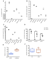
Figure 1. Increased IL-6 expression after chemotherapy associated with cancer progression and poor clinical outcome.
(A) Mean of IL-6 expression (reads per kilobase of transcript, per million mapped reads; RPKM) in patients with free tumor survival duration less than 12 months and more than 12 months after initial chemotherapy. Data was drawn from TCGA ovarian cancer portal. There are 20 patients with free tumor survival duration less than 12 months and 22 patients with free tumor survival duration more than 12 months after initial chemotherapy. (B) Survival probability of patients with high-grade serous ovarian cancer correlates with IL-6 expression levels in tumors after chemotherapy (P = 0.0023).
Figure 2
Figure 2. Platinum-induced IL-6 secretion contributes to enrichment of OCSC in residual tumors.
(A) Basal expression of IL-6 and (B) IL-6 receptor mRNA expression in human OC cells (ng/ml) were measured by qPCR. Bars represent average measurements of 3 independent experiments ± SD (n = 3). (C) COV318, OVCAR4, Kuramochi, EFO-27, HeyA8, A2780_CR5, SKOV3, and A2780 OC cells were cultured under starving condition for 24 hours. ELISA was used to measure IL-6 levels in the conditioned media. IL-6 secretion (pg/ml/1 × 105 cells) was determined and normalized to the cell number (n = 3). (D) Kuramochi, OVCAR4, and A2780 OC cells were cultured under starving conditions for 24 hours and then treated with CDDP (IC50 or half of IC50). ELISA was used to measure IL-6 levels in the CM. IL-6 secretion (pg/ml/ 1 × 10⁵ cells) was determined and normalized to the cell number. Average fold change of IL-6 secretion (± SD) of CDDP-treated cells compared with control-treated is shown (2-tailed Student’s t test, *P < 0.05, **P < 0.01, and ***P < 0.001) (n = 3). (E) Nude mice were bearing A2780 i.p.-derived xenograft tumors were treated with carboplatin (50 mg/kg, weekly for 3 weeks). Blood samples and xenograft tumors were collected after CDDP treatment. Average relative IL-6 expression level (± SEM) in the plasma compared baseline plasma IL-6 level was measured by IL-6 ELISA and shown (n = 6). (F) IL-6 and IL-6R expression in the tumor residuals treated with control and carboplatin were measured by using qPCR. Mean fold change of IL-6 and IL-6R expression of 6 independent experiments is reported. Two-tailed Student’s t test was used to analyze statistical significance (*P < 0.05, **P < 0.01, and ***P < 0.001).
Figure 3
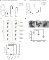
Figure 3. Upregulation of the IL-6 signaling pathway in ALDH+ ovarian cancer stem cells.
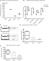
(A) Five thousand ALDH+/– cells were FACS sorted from Kuramochi, OVCAR4, and A2780 cells into 96-well plates. IL-6 levels were measured by IL-6 ELISA at 24 hours after starving conditions. Average relative IL-6 secretion levels (± SD) of ALDH+ OCs compared with ALDH cells is shown (n = 3). (B) Expression of ALDH1A1, IL-6, and IL-6R were measured by qPCR in Kuramochi-, A2780-, and OVCAR4-derived ALDH+ cells, compared with respective ALDH cells. Average fold change (± SD) of 3 independent experiments is shown. (*P < 0.05, **P < 0.01, and ***P < 0.001) (n = 3). (C) Kuramochi cells were transfected with scrambled shRNA, shALDH1A1, or shIL-6 RNA. Side scatter of FACS analysis of the percentage of ALDH+ cell population in Kuramochi cells with stable ALDH1A1 and IL-6 knockdown expression. Average percentage of ALDH+ cells (± SD) is shown below (n = 3, 1-way ANOVA, *P < 0.05). (D) Representative pictures of spheroids formed by 500 shcontrol, shALDH1A1, and shIL-6 knockdown Kuramochi cells, which were cultured in stem cell culture condition for 14 days; the average number of spheroids (± SD) is shown below (n = 3, 1-way ANOVA, *P < 0.05). Scale bar: 100 μm.
Figure 4
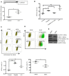
Figure 4. CDDP-induced IL-6 regulates ALDH1A expression and OCSC enrichment.
(A) OC-derived (Kuramochi-derived) ALDH cells were cotransfected with pGL3-ALDH1A1-Luc and renilla luciferase plasmid vector (pRL) or pGL3-Luc and pRL. Transfected cells were cultured with starving medium for 24 hours and were then treated with IL-6 (100 ng/ml). Luciferase signals recorded 3 hours after drug treatment. Renilla luciferase activity used for normalization. Average fold changes (± SD) of relative luciferase unit (RLU) compared with pGL3 are shown (n = 3). (B) Quantification of FACS analysis of the percentage of ALDH+ in Kuramochi OC cells treated with CDDP (3 μM, 3 hours), CDDP+IL-6-Nab (1 μg/ml) and IL-6 (500 ng/ml) for 72 hours. Average percentage of ALDH+ cells (± SD) is shown on the graph, and the quantification is shown (n = 3, 1-way ANOVA, *P < 0.05 and ***P < 0.001). (C) Side scatter of FACS analysis of percentage of ALDH+ cells population in Kuramochi-derived ALDH cells, which were treated with CDDP (IC50, weekly for 3 weeks) and IL-6 (100 ng/ml, weekly for 3 weeks). Average percentage of ALDH+ cells (± SD) is shown on the graph, and the quantification is shown (n = 3, 1-way ANOVA, **P < 0.01 and ***P < 0.001). (D) FACS analysis of the percentage of Kuramochi-derived ALDH+ cells treated daily with DMSO or Stattic (3 μM) for 3 days. Average percentage of ALDH+ cells ± SD is shown on the graph (n = 3). Two-tailed Student’s t test was used to analyze statistical significance (*P < 0.05). (E) Protein expression of ALDH1A1, DNMT1, pSTAT3, STAT3, and GAPDH in DMSO or Stattic-treated Kuramochi_ALDH+ cells were determined by Western blot (n = 2).
Figure 5
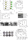
Figure 5. IL-6-Nab–guadecitabine combination eliminates ALDH+ cells by inhibiting ALDH1A1 expression.
(A) FACS analysis of the percentage of Kuramochi-derived ALDH+ cells treated daily with guadecitabine (100 nM), IL-6-Nab (1 μg/ml), or IL-6-Nab– guadecitabine for 4 days. Average percentage of ALDH+ cells ± SD is shown on the side scatter graph (left), and the quantification is shown (right) (n = 3, 1-way ANOVA). (B) Average fold change of ALDH1A1 expression ± SD in Kuramochi-derived ALDH+ cells treated with guadecitabine, IL-6-Nab, or IL-6-Nab–guadecitabine compared with control cells was determined by qPCR (n = 3, 1-way ANOVA, *P < 0.05 and ***P < 0.001) (C) Kuramochi-derived ALDH+ cells were treated daily with guadecitabine (100 nM), IL-6-Nab (1 μg/ml), or IL-6-Nab–guadecitabine for 4 days. The protein expression of ALDH1A1, DNMT1, pSTAT3, STAT3, and GAPDH was determined by western blot (n = 2). (D) Spheroid formation assay of 500 conditioned Kuramochi cells, which were treated with control, IL-6-Nab (400 ng/ml), and guadecitabine (100 nM, 3 days) or in combination. Cells were directly sorted in 96 low-attached plates and cultured in stem cell condition for 14 days. Representative images and the average number of spheroids ± SD are shown in the graph. Scale bar: 100μm (n = 3, 1-way ANOVA, *P < 0.05). (E) Kuramochi_ALDH+ cells were treated daily with guadecitabine (100 nM), IL-6-Nab (1 μg/ml), or IL-6-Nab– guadecitabine for 4 days. Five hundred pretreated cells were reseeded into 6-well plates for clonogenic survival assays. Representative images and the average number and of colonies formed by pretreated Kuramochi_ALDH+ cells with the conditions described previously and pretreated Kuramochi_ALDH+ cells after exposure to cisplatin (3 μM) for 3 hours (n = 3, 2-way ANOVA, Dunnett’s and Sidak’s multiple comparisons tests used for multiple comparison, *P < 0.05, **P < 0.01, ***P < 0.001, and ****P < 0.0001).
Figure 6
Figure 6. Combination of IL-6 neutralizing antibody with guadecitabine as maintenance therapy reduces ALDH+ population in platinum-treated tumor residuals.
(A) Schematic diagram of experimental design including carboplatin treatment phase followed by randomization to vehicle, guadecitabine, IL-6-Nab or the drug combination. (B–D) Effects of different treatment strategies on total tumor weight (n = 5, 1-way ANOVA, *P < 0.05 and ***P < 0.001) (B), total number of tumor metastatic sites (n = 5, 1-way ANOVA, *P < 0.05) (C), and average of the percentage of ALDH+ cells in xenografts (n = 5, 1-way ANOVA, ***P < 0.001) (D). (E) The average number ± SD of spheroids formed by 10,000 cells dissociated from xenografts, which were treated with vehicle, carboplatin, carboplatin + vehicle (Carbo_resi), carboplatin + guadecitabine, carboplatin + IL-6-Nab, or carboplatin + guadecitabine + IL-6-Nab (n = 5, 1-way ANOVA, **P < 0.01 and ***P < 0.001). (F) mRNA expression of ALDH1A1, Sox2, Bmi1, IL-6, and IL-6 receptor (n = 3, 1-way ANOVA, *P < 0.05, **P < 0.01, and ***P < 0.001).
Figure 7
Figure 7. Normal omental fibroblasts (NOFs) enhance IL-6 secretion.
(A) Baseline IL-6 secretion level by Kuramochi, OVCAR4, A2780, and NOFs (n = 3). (B) NOFs cells were cultured alone or cocultured with OCs (Kuramochi + NOFs, OVCAR4 + NOFs, A2780 + NOFs) under starving condition for 24 hours and then treated with CDDP (IC50 or half of IC50). IL-6 levels in the conditioned media were measured at 24 hours after exposure to CDDP by ELISA. IL-6 secretion (pg/ml/1 × 105 cells) was determined and normalized to the cell number. Average relative IL-6 secretion (± SD) of CDDP-treated cells compared with control-treated is shown in the graph (n = 3, 2-tailed Student’s t test, *P < 0.05, **P < 0.01, and ***P < 0.001). (C) Schematic diagram of transwell coculture system. In this 0.45-μm transwell coculture system, OC cells (Kuramochi_ALDH/A2780_ALDH cells) with starving medium (DMEM supplemented with 0.5% FBS) were seeded in the upper chambers alone (top) or with NOFs, which were seeded on the bottom chamber in DMEM supplemented with 0.5% FBS (bottom). (D and E) Average fold change with range of mRNA ALDH1A1, Sox2, and Bmi1 expression in (D) Kuramochi- and (E) A2780-derived ALDH cells cocultured with NOFs compared with respective ALDH cells alone shown in the graph (average fold change ± SD). Two-tailed Student’s t test was used to analyze statistical significance (n = 3, *P < 0.05, **P < 0.01, and ***P < 0.001).

References

    1. Berek JS, et al. Advanced epithelial ovarian cancer: 1998 consensus statements. Ann Oncol. 1999;10 Suppl 1:87–92. - PubMed
    1. Bowtell DD, et al. Rethinking ovarian cancer II: reducing mortality from high-grade serous ovarian cancer. Nat Rev Cancer. 2015;15(11):668–679. doi: 10.1038/nrc4019. - DOI - PMC - PubMed
    1. Dean M, Fojo T, Bates S. Tumour stem cells and drug resistance. Nat Rev Cancer. 2005;5(4):275–284. doi: 10.1038/nrc1590. - DOI - PubMed
    1. Wang Y, et al. Epigenetic targeting of ovarian cancer stem cells. Cancer Res. 2014;74(17):4922–4936. doi: 10.1158/0008-5472.CAN-14-1022. - DOI - PMC - PubMed
    1. Zhang S, et al. Identification and characterization of ovarian cancer-initiating cells from primary human tumors. Cancer Res. 2008;68(11):4311–4320. doi: 10.1158/0008-5472.CAN-08-0364. - DOI - PMC - PubMed

Publication types

MeSH terms