. 2018 Dec 5;9(1):5201.
doi: 10.1038/s41467-018-07511-4.

NSD2 is a conserved driver of metastatic prostate cancer progression

Affiliations

NSD2 is a conserved driver of metastatic prostate cancer progression

Alvaro Aytes et al. Nat Commun. .

Abstract

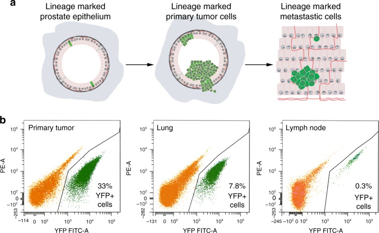
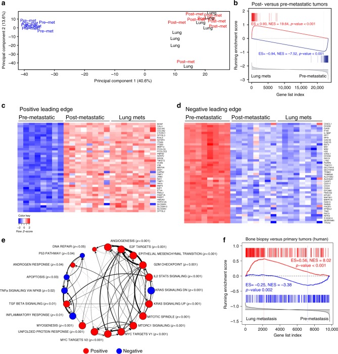
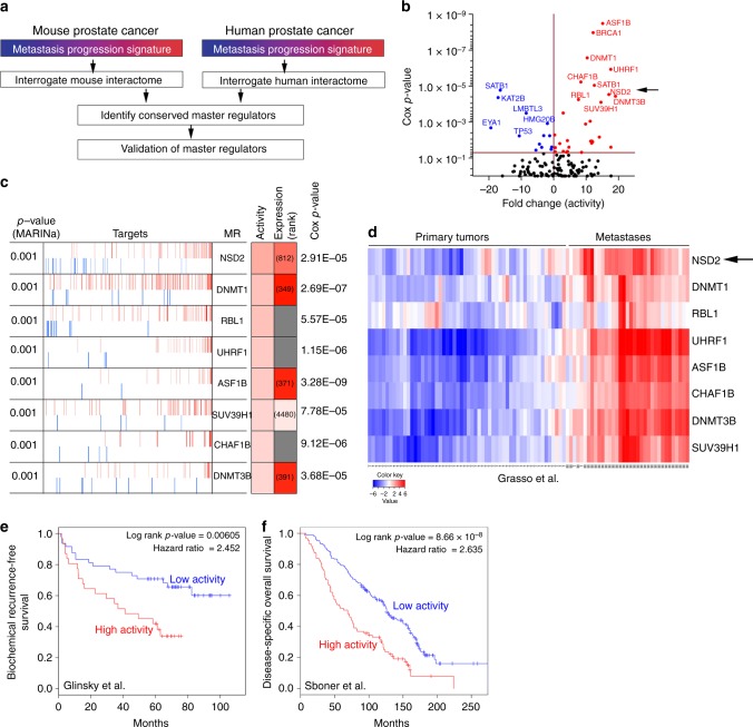
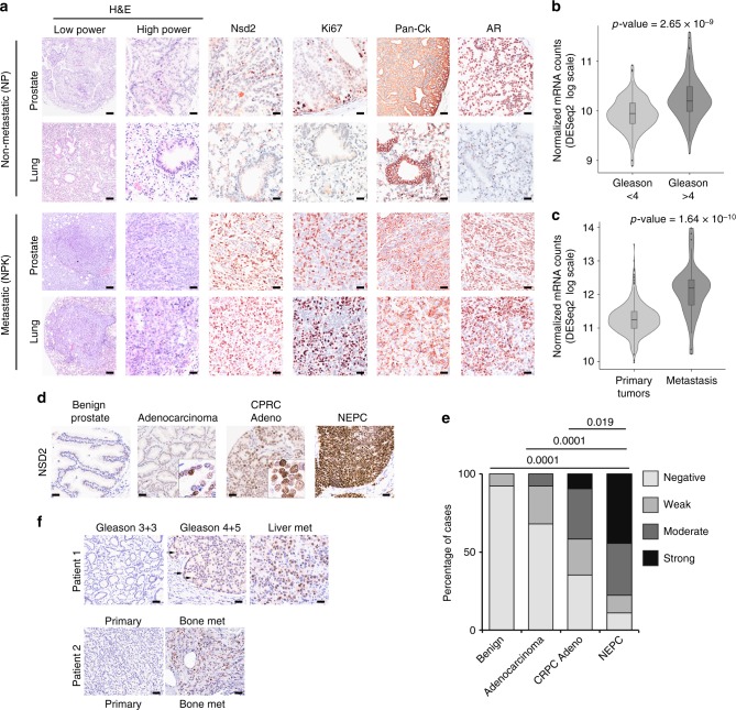
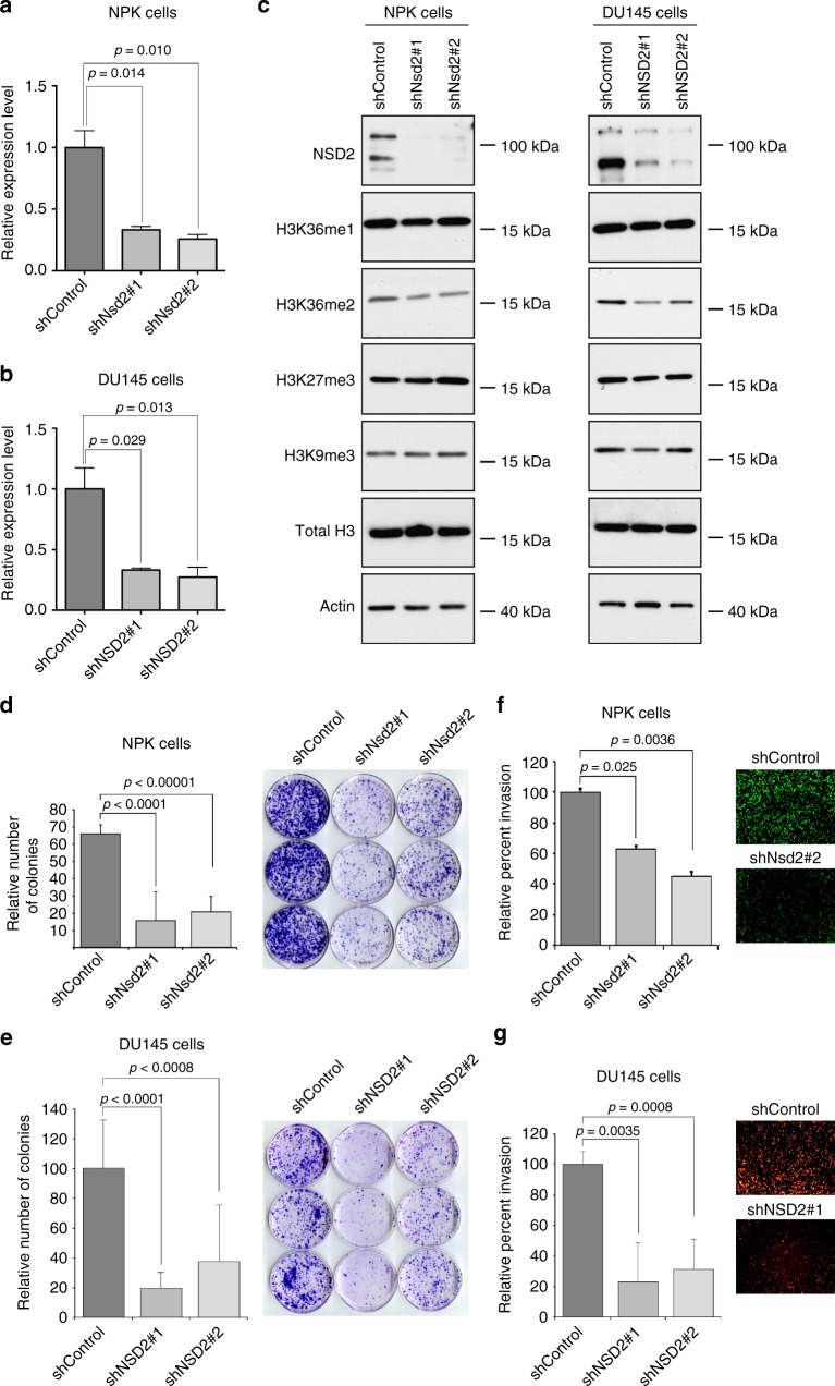
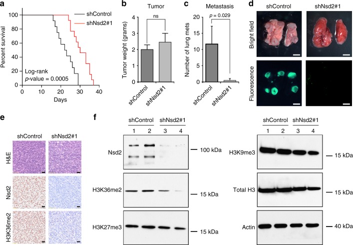
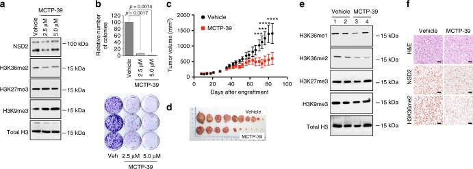
Deciphering cell-intrinsic mechanisms of metastasis progression in vivo is essential to identify novel therapeutic approaches. Here we elucidate cell-intrinsic drivers of metastatic prostate cancer progression through analyses of genetically engineered mouse models (GEMM) and correlative studies of human prostate cancer. Expression profiling of lineage-marked cells from mouse primary tumors and metastases defines a signature of de novo metastatic progression. Cross-species master regulator analyses comparing this mouse signature with a comparable human signature identifies conserved drivers of metastatic progression with demonstrable clinical and functional relevance. In particular, nuclear receptor binding SET Domain Protein 2 (NSD2) is robustly expressed in lethal prostate cancer in humans, while its silencing inhibits metastasis of mouse allografts in vivo. We propose that cross-species analysis can elucidate mechanisms of metastasis progression, thus providing potential additional therapeutic opportunities for treatment of lethal prostate cancer.


Conflict of interest statement

M.A.R. receives research support from Jansen, Eli Lilly, and Sanofi-Aventis; is a co-inventor on gene fusion prostate cancer for diagnostic and therapeutic uses, and is a co- founder Thucydx, LLC. A.A. and L.P. receive support from Roche Pharma and Astellas Pharma directed to support the ProCURE research program. A.C. is a founder, equity holder, and serves on the advisory board of DarwinHealth Inc. The remaining authors declare no competing interests.

Figures

Fig. 1
Fig. 1
Strategy for molecular profiling of tumors and metastases. a Lineage tracing of YFP-labeled (green) prostate epithelial cells at the time of tumor induction leads to YFP-labeled cells in tumors and metastases. b Lineage-marked cells from primary tumors, or lung or lymph node metastases were isolated by fluorescence activated cell sorting (FACS). Shown are representative images with percentages of YFP-labeled cells indicated; axes show fluorescent intensity of the fluorescein isothiocynate (FITC-A) and phycoeritrin (PE) channels
Fig. 2
Fig. 2
A molecular signature of de novo metastasis progression. a Principal component analysis (PCA) comparing expression profiles from pre-metastatic (pre-met, blue) or post-metastatic (post-met, red) primary tumors, or lung metastases (lung, black) from NPK mice (n = 8/group). Note that Principal Component 1, capturing 40.6% of gene expression variance, effectively distinguishes pre-metastatic tumors from post-metastatic tumors and lung metastases. b GSEA comparing a reference signature of mouse lung metastases (lung mets) versus pre-metastatic tumors to a query signature of mouse post-metastatic versus pre-metastatic tumors. c, d Heat map representations of differentially expressed genes from the positive and negative leading edges, respectively, from the GSEA in panel b. The color key shows relative expression levels of the differentially expressed genes (red corresponds to overexpressed genes while blue corresponds to underexpressed genes). e Pathway enrichment analysis using the mouse metastasis progression signature defined between lung metastasis versus pre-metastatic tumors, as in panel b, to query the hallmark pathways dataset from the molecular signatures database (MSigDB). Red and blue nodes indicate positive and negative enrichment, respectively (p < 0.05). Thickness of arrows indicate the overlap of genes in the leading edges. The p-values correspond to the GSEA enrichment, and the relative size of the node indicates the relative p-value, as shown. f Cross-species GSEA comparing a reference mouse metastasis progression signature (lung metastasis versus pre-metastatic tumors, as in panel b) with a query gene set from a human metastasis signature defined between bone metastasis biopsies versus primary tumors from Balk et al. (Supplementary Table 1). For GSEA, red vertical bars indicate overexpressed query genes and blue vertical bars indicate underexpressed query genes. GSEA were done using the top 200 differentially expressed genes; p-values were calculated using 1000 gene permutations. ES: enrichment score, NES: normalized enrichment score
Fig. 3
Fig. 3
Conserved master regulators of metastasis progression. a Strategy: mouse and human metastasis progression signatures (as in Fig. 2f) were used to interrogate mouse and human prostate cancer interactomes, respectively, using the MARINa algorithm. Independent lists of mouse and human master regulators (MRs) were integrated to identify conserved MRs, which were prioritized by clinical and functional validation. b Scatter plot showing the association of the 136 conserved MRs (Supplementary Data 3) to clinical outcome using the Sboner et al. dataset, which reports prostate cancer-specific survival as the clinical endpoint (Supplementary Table 1). The Y axis represents the Cox proportional hazard p-value and the X axis represents the fold change based on MR activity. MRs that are inactive (blue) relative to primary tumors have negative fold change values and those that are active (red) have positive fold change values. c Summary of the 8 candidate MRs depicting their positive (activated; red bars) and negative (repressed; blue bars) targets. Shaded boxes show the ranks of differential activity and differential expression (darker is higher and lighter is lower); the numbers indicate their rank in the differential expression signature (gray indicates that a specific gene is not present on the utilized gene expression platform, yet its targets are present). P-values for Cox proportional hazard were estimated using a Wald test based on time to prostate cancer-specific death in Sboner et al. d Heatmap showing hierarchical clustering of primary tumors and metastasis from the Grasso et al. cohort (Supplementary Table 1) based on the activities of the 8 candidate MRs. The color key shows activity levels of MRs (i.e., NESs), where red corresponds to increased activity and blue correspond to decreased activity of the MRs. e, f Kaplan–Meier survival analysis based on the activity levels of the 8 candidate MRs in: e Glinsky et al. (n = 79), with biochemical recurrence as the clinical end-point; and f Sboner et al. (n = 281), with prostate cancer-specific survival as the clinical endpoint (Supplementary Table 1). P-values were estimated using a log-rank test to determine the difference in outcomes between patients with higher MR activity levels (red) versus those with lower/no MR activity (blue)
Fig. 4
Fig. 4
Expression of NSD2 in prostate cancer and metastases. a Immunostaining of Nsd2 and other markers on mouse primary tumors and metastases. Shown are representative H&E images and immunostaining for the indicated antibodies from non-metastatic NP mice and metastatic NPK mice (n = 4/group). Scale bars in the low power H&E images represent 100 microns, and all other panels 50 microns. b, c Violin plots comparing mRNA expression levels of NSD2 in TCGA and SU2C human prostate cancer cohorts (Supplementary Table 1). b compares primary tumors from TCGA divided based on pathological grade [Gleason <4 (n = 104) or ≥ 4 (n = 173)], as indicated. c compares primary tumors and metastases from a cohort combining primary tumors from TCGA (all Gleason scores; n = 333) and metastases from SU2C (n = 51). P-values were estimated using the two-sample two-tailed Welch t-test. d, e Immunostaining of NSD2 on a human prostate tissue microarray (TMA) (n = 100 independent cases). d shows representative images representing benign prostate, untreated localized adenocarcinoma, castration-resistant adenocarcinoma (CRPC-Adeno) and neuroendocrine prostate cancer (NEPC). Nuclear staining intensity was evaluated blinded by a pathologist and scored as negative (or present in <5% of nuclei), weak, moderate or strong. Scale bars represent 50 microns. e shows quantification of nuclear intensity staining for each score (negative, weak, moderate, and strong). The p-values compare negative/weak staining versus moderate/strong staining in each group and were calculated using a two-tailed Fisher's exact test. f Immunostaining of NSD2 on matched patient sets of primary prostate cancer and distant metastasis to soft tissues or bone, as indicated. Patient 1 shows representative images of lower pathological grade (Gleason 3 + 3), which is negative for NSD2, and higher pathological grade (Gleason 4 + 5) and a liver metastasis that have increasing expression of NSD2. Patient 2 shows a high grade primary tumor (primary) that is negative for NSD2 and a matched bone metastasis in which NSD2 staining is readily detected. Scale bars represent 50 μ
Fig. 5
Fig. 5
Silencing of NSD2 abrogates tumorigenicity in vitro. Panels a-g show in vitro analyses of NSD2 silencing in a mouse metastatic cell line (NPK cells) and a human advanced prostate cancer cell line (DU145 cells). Cells were infected with control shRNA or two independent shRNAs for mouse or human NSD2, respectively. a, b Validation of NSD2 silencing in NPK and DU145 cells, as indicated, using quantitative real-time PCR (qPCR). c Western blot analyses of NSD2-silenced or control NPK and DU145 cells, as indicated, showing reduced expression of NSD2, which is accompanied by reduction of the H3K36me2 mark, but not the H3K36m1 or the other histone marks shown. The position of a molecular marker is shown; uncropped images are provided in Supplementary Figure 5. d, e Colony formation assays in NPK and DU145 cells, as indicated showing quantification (left) representative images (right). f, g Invasion assays in NPK and DU145 cells, as indicated showing quantification (left) and representative images (right). Experiments were done in three independent biological replicates each in triplicate; p-values were calculated using a two-tailed Student's t-test. Error bars represent the standard deviation (s.d.) from the mean
Fig. 6
Fig. 6
Silencing of Nsd2 abrogates metastasis in vivo. Panels af show in vivo analyses of Nsd2 silencing in a mouse metastatic cell line (NPK cells). Cells (1 × 106 cells) were engrafted subcutaneously into the flank of nude mice and the mice were monitored for up to 40 days. Studies were done using 2 independent shRNA for Nsd2; representative data for shRNA#1 is shown. a Survival analyses with the endpoint being tumor volume of 1.5 cm3 (n = 10/group). The p-value was calculated using a log-rank test. b Analyses of tumor weights (in grams) at the time of killing (total n = 9/group). c Number of lung metastases per mouse (total n = 9/group). b, c p-values were calculated using the Mann–Whitney U test; error bars represent the standard deviation (s.d.) from the mean. d Representative whole mount and epifluorescence images of lung metastases. Scale bar represent 100 microns. e Representative immunostaining of shControl and shNsd2 tumors using the indicated antibodies (n = 4/group). Scale bars represent 50 μ. f Western blot analysis showing representative cases from the shControl (lanes 1, 2) and shNsd2 (lanes 3, 4) tumors using the indicated antibodies (total n = 4/group). The position of a molecular marker is shown; uncropped images are provided in Supplementary Figure 5
Fig. 7
Fig. 7
Pharmacological treatment. a, b Pharmacological treatment in vitro. DU145 cells were treated with MCTP-39 at the indicated concentrations for 72 h. Panel a shows western blot data using the indicated antibodies. The position of a molecular marker is shown; uncropped images are provided in Supplementary Figure 6. b depicts colony forming assays showing quantification (top) and representative images (bottom). Shown are representative data from 3 independent experiments, each done in triplicate. Error bars represent the standard deviation (s.d.) from the mean; p-values were calculated using a two-tailed Student's t-test. cf Pharmacological treatment in vivo. DU145 cells (5 × 106 cells) were engrafted subcutaneously into male nude mouse hosts. After 1 week of growth, the tumor-bearing mice were randomized by cage to the vehicle (black) or MCTP-39 (red) treatment groups and treated with 10 mg/kg with MCTP-39 (or vehicle only) for up to 3 months. Tumor volume was monitored using calipers and calculated using the formula [Volume = (width)2 x length/2]. Total mice analyzed for vehicle were 14 and for MCTP-39-treatment were 15 in two independent experiments. c Tumor volume. Two-way analysis of variance (ANOVA) was used to calculate the significance (p-value) of the difference between the vehicle and treatment group; ***p< 0.001 and ****p < 0.0001. d Representative tumors collected at at the time of euthanasia. e Western blot showing 2 examples from vehicle (lanes 1, 2) and MCTP-39 (lanes 3, 4) treated tumors using the indicated antibodies (total n = 4/group). The position of a molecular marker is shown; uncropped images are provided in Supplementary Figure 6. f Representative immunostaining for NSD2 and H3k36me3 from vehicle and MCTP-39 treated mice (n = 4/group). Scale bars represent 50 μ

References

    1. Lambert AW, Pattabiraman DR, Weinberg RA. Emerging biological principles of metastasis. Cell. 2017;168:670–691. doi: 10.1016/j.cell.2016.11.037. - DOI - PMC - PubMed
    1. Vanharanta S, Massague J. Origins of metastatic traits. Cancer Cell. 2013;24:410–421. doi: 10.1016/j.ccr.2013.09.007. - DOI - PMC - PubMed
    1. Bos PD, Nguyen DX, Massague J. Modeling metastasis in the mouse. Curr. Opin. Pharmacol. 2010;10:571–577. doi: 10.1016/j.coph.2010.06.003. - DOI - PMC - PubMed
    1. Maddipati R, Stanger BZ. Pancreatic cancer metastases harbor evidence of polyclonality. Cancer Discov. 2015;5:1086–1097. doi: 10.1158/2159-8290.CD-15-0120. - DOI - PMC - PubMed
    1. Aytes A, et al. ETV4 promotes metastasis in response to activation of PI3-kinase and Ras signaling in a mouse model of advanced prostate cancer. Proc. Natl Acad. Sci. USA. 2013;110:E3506–E3515. doi: 10.1073/pnas.1303558110. - DOI - PMC - PubMed

Publication types

MeSH terms