. 2018 Dec 5;8(1):17654.
doi: 10.1038/s41598-018-36182-w.

Extracellular Protein Fibulin-7 and Its C-Terminal Fragment Have In Vivo Antiangiogenic Activity

Affiliations

Extracellular Protein Fibulin-7 and Its C-Terminal Fragment Have In Vivo Antiangiogenic Activity

Tomoko Ikeuchi et al. Sci Rep. .

Abstract

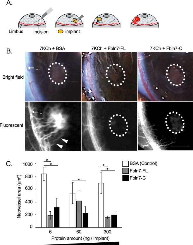
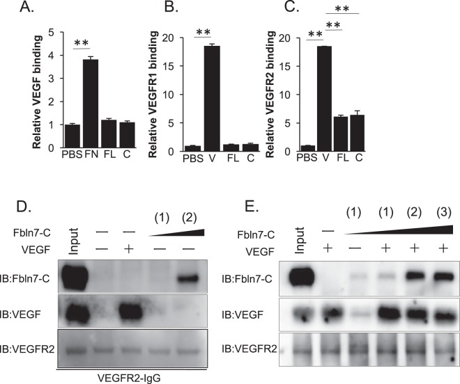
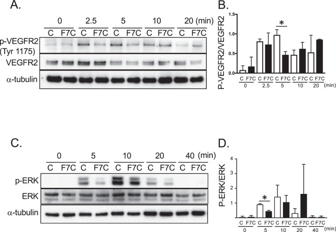
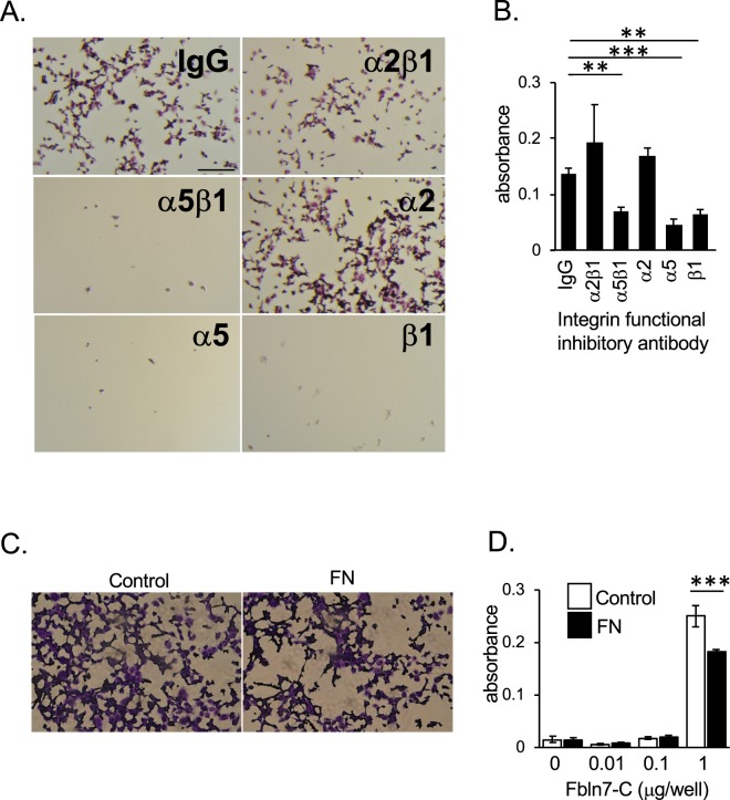
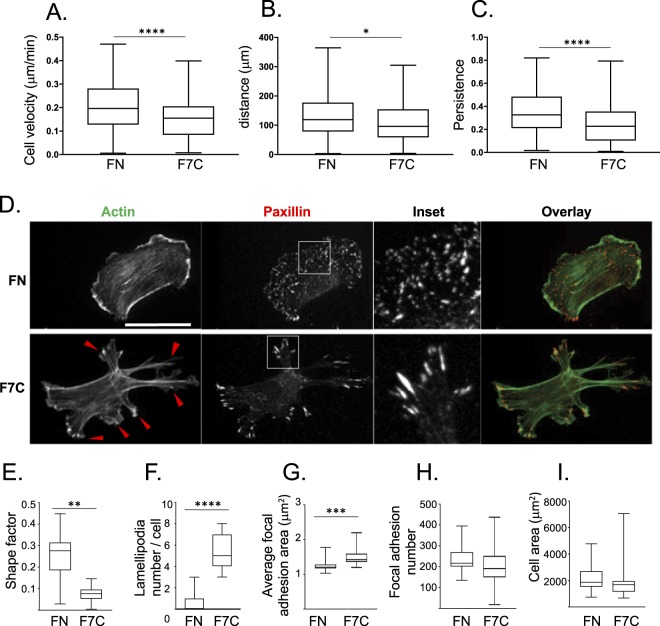
Angiogenesis is crucial for tissue development and homeostasis; however, excessive angiogenesis can lead to diseases, including arthritis and cancer metastasis. Some antiangiogenic drugs are available, but side effects remain problematic. Thus, alternative angiogenesis inhibition strategies are needed. Fibulin-7 (Fbln7) is a newly discovered member of the fibulin protein family, a group of cell-secreted glycoproteins, that functions as a cell adhesion molecule and interacts with other extracellular matrix (ECM) proteins as well as cell receptors. We previously showed that a recombinant C-terminal Fbln7 fragment (Fbln7-C) inhibits tube formation by human umbilical vein endothelial cells (HUVECs) in vitro. In the present study, we examined the in vivo antiangiogenic activity of recombinant full-length Fbln7 (Fbln7-FL) and Fbln7-C proteins using a rat corneal angiogenesis model. We found that both Fbln7-FL and Fbln7-C inhibited neovascularization. Fbln7-C bound to vascular endothelial growth factor receptor 2 (VEGFR2), inhibiting VEGFR2 and ERK phosphorylation and resulting in reduced HUVEC motility. HUVEC attachment to Fbln7-C occurred through an interaction with integrin α5β1 and regulated changes in cellular morphology. These results suggest that Fbln7-C action may target neovascularization by altering cell/ECM associations. Therefore, Fbln7-C could have potential as a therapeutic agent for diseases associated with angiogenesis.


Conflict of interest statement

The authors declare no competing interests.

Figures

Figure 1
Figure 1
Fbln7-FL and Fbln7-C inhibit 7KCh-induced neovascularization in vivo. (A) Surgical procedure for the rat corneal angiogenesis model. (B) Rat corneal angiogenesis model. Implants containing 300 ng protein/implant were placed into the anterior chamber, and fluorescent images were taken 10 days post-implantation. Three different implants were inserted: 7KCh and BSA (control), 7KCh and Fbln7-FL, and 7KCh and Fbln7-C. White dotted circles indicate implants. White triangles indicate neovasculature from limbus towards the implant. L: Limbus; triangles indicate neovasculature; scale bar: 1 mm. (C) Protein dose-dependent experiments in the rat corneal angiogenesis model. Neovessel area was calculated for each experiment, with 6, 60, and 300 ng protein in the implants. N = 6, *P < 0.05.
Figure 2
Figure 2
Fbln7 and Fbln7-C bind to VEGFR2, but not to VEGFR1 or VEGF. Binding between Fbln7-FL/Fbln7-C and (A) VEGF, (B) VEGFR1, or (C) VEGFR2 by ELISA. FN: Fibronectin, FL: Fbln7-FL, C: Fbln7-C, V: VEGF. **P < 0.01. (D) Binding between Fbln7-C and VEGFR2 by pull-down assays. (E) Binding between Fbln7-C and VEGFR2 at various protein ratios: (1) VEGFR2:Fbln7-C = 1:1; (2) VEGFR2:Fbln7-C = 1:10; (3) VEGFR2:Fbln7-C = 1:20. All samples included the VEGFR2 protein. Each row is a different gel, but every gel was loaded with the same sample in equal volumes.
Figure 3
Figure 3
Fbln7-C inhibits VEGFR2 phosphorylation and ERK phosphorylation in the VEGF-VEGFRs signaling pathway. (A,B) VEGFR2 Tyr1175, and (C,D) ERK phosphorylation levels with Fbln7-C (100 µg/ml) pretreatment and after VEGF (5 ng/ml) stimulation were confirmed by western blotting. C: control, F7C: Fbln7-C *P < 0.05. All samples included the VEGFR2 protein. Each row is a different gel, but every gel was loaded with the same sample in equal volumes.
Figure 4
Figure 4
Integrin α5β1 is involved in HUVEC attachment to Fbln7-C. HUVEC attachment to Fbln7-C (1 µg/well)-coated wells with treatment by antibody integrin functional inhibitors or control IgG. (A) Brightfield images of HUVECs treated with integrin functional inhibitors. Scale bar: 1 mm. (B) Quantification of HUVEC attachment to Fbln7-C-coated wells with integrin functional inhibitor treatment by measuring CCK8 absorbance. ***P < 0.0001, **P < 0.01. (C,D) HUVEC attachment competition assays between fibronectin and Fbln7-C. Numbers of attached cells were determined by CCK8 staining. (C) Inhibitory competition of HUVEC binding to a Fbln7 substrate by soluble fibronectin. Fbln7-C-coated well (1 µg/well) with and without pre-treatment by fibronectin. FN: Fibronectin. (D) Quantification of HUVEC attachment to Fbln7-C-coated wells with fibronectin treatment. HUVECs were plated on dishes coated with different amounts of Fbln7 and cultured with or without fibronectin. After 30 minutes culture, numbers of attached cells were measured by CCK8 staining. ***P < 0.0001.
Figure 5
Figure 5
Fbln7-C affects focal adhesion area and actin filaments to inhibit cell motility. (AC) Cell motility on fibronectin or Fbln7-C-coated dishes stimulated with VEGF and analyzed using time-lapse imaging. Cell motility is evaluated by (A) cell velocity, (B) distance, (C) and persistence. N = 3 n > 150, ****P < 0.0001, **P < 0.01, *P < 0.01. (DI) Cell morphology differences between cells on fibronectin and Fbln7-C-coated dishes evaluated by staining for focal adhesion sites (paxillin; red) and actin filaments (phalloidin; green). (D) Immunofluorescence staining. Cell shape is evaluated by (E) shape factor and (F) number of lamellipodia. Focal adhesion area is evaluated by (G) average focal adhesion area, (H) focal adhesion number and (I) cell area. FN: Fibronectin, F7C:Fibulin7-C, scale bar: 100 μm, N = 3 n > 80, ****P < 0.0001, **P < 0.01, *P < 0.01.

References

    1. Takahashi HSM. The vascular endothelial growth factor (VEGF)/VEGF receptor system and its role under physiological and pathological conditions. Clin Sci (Lond). 2005;109:227–241. doi: 10.1042/CS20040370. - DOI - PubMed
    1. Kamba T, McDonald DM. Mechanisms of adverse effects of anti-VEGF therapy for cancer. British Journal of Cancer. 2007;96:1788–1795. doi: 10.1038/sj.bjc.6603813. - DOI - PMC - PubMed
    1. Clamp AR, Jayson GC. The clinical potential of antiangiogenic fragments of extracellular matrix proteins. Br J Cancer. 2005;93:967–972. doi: 10.1038/sj.bjc.6602820. - DOI - PMC - PubMed
    1. Hombrebueno JR, Ali IHA, Xu H, Chena M. Sustained intraocular VEGF neutralization results in retinal neurodegeneration in the Ins2Akita diabetic mouse. Sci Rep. 2015;5:18316. doi: 10.1038/srep18316. - DOI - PMC - PubMed
    1. Folkman J. Endogenous angiogenesis inhibitors. APMIS: acta pathologica, microbiologica, et immunologica Scandinavica. 2004;112:496–507. doi: 10.1111/j.1600-0463.2004.apm11207-0809.x. - DOI - PubMed

Publication types

MeSH terms

Substances