Review
. 2019 May;17(5):321-328.
doi: 10.1038/s41579-018-0120-2.

Prisoners of war - host adaptation and its constraints on virus evolution

Affiliations
Review

Prisoners of war - host adaptation and its constraints on virus evolution

Peter Simmonds et al. Nat Rev Microbiol. 2019 May.

Abstract

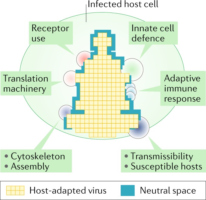
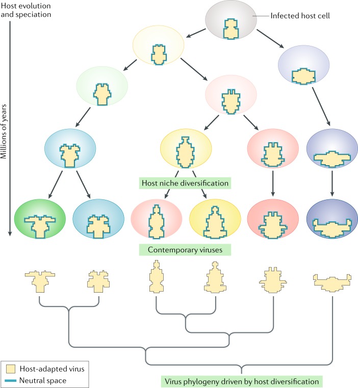
Recent discoveries of contemporary genotypes of hepatitis B virus and parvovirus B19 in ancient human remains demonstrate that little genetic change has occurred in these viruses over 4,500-6,000 years. Endogenous viral elements in host genomes provide separate evidence that viruses similar to many major contemporary groups circulated 100 million years ago or earlier. In this Opinion article, we argue that the extraordinary conservation of virus genome sequences is best explained by a niche-filling model in which fitness optimization is rapidly achieved in their specific hosts. Whereas short-term substitution rates reflect the accumulation of tolerated sequence changes within adapted genomes, longer-term rates increasingly resemble those of their hosts as the evolving niche moulds and effectively imprisons the virus in co-adapted virus-host relationships. Contrastingly, viruses that jump hosts undergo strong and stringent adaptive selection as they maximize their fit to their new niche. This adaptive capability may paradoxically create evolutionary stasis in long-term host relationships. While viruses can evolve and adapt rapidly, their hosts may ultimately shape their longer-term evolution.


Conflict of interest statement

The authors declare no competing interests.

Figures

Fig. 1
Fig. 1. Virus genome nucleotide substitution rates of different observation periods.
Plots of substitution rates of DNA and RNA viruses calculated over different time periods using different methods are shown. These include Bayesian evolutionary reconstructions and rates inferred from instances of virus–host co-evolution (see the figure key). Data used in the figure are based on a previous analysis of published virus substitution rates with different genomic configurations and expanded with more recent published data (listed in full in Supplementary information). Three groups are depicted: double-stranded DNA (dsDNA) viruses in Baltimore group I (part a), single-stranded DNA (ssDNA) viruses in Baltimore group II (part b) and reverse transcribing (RT) viruses in Baltimore groups VI and VII (part c). These groups showed a remarkably similar relationship between substitution rate (y axis) and observation times over which substitution rates were calculated (plotted on a log-transformed scale on the x axis) despite their intrinsic differences in replication error rates and evolutionary histories. The regression line is based on substitution rates calculated from co-evolution and phylogeny methods. Rates inferred from very ancient co-evolutionary scenarios among RT viruses show a potential flattening of substitution rates as they approach those of host genes (mean value 2.2 × 10⁻⁹ substitutions per site per year (SSY)). Evolutionary rates estimated from ancient DNA (aDNA) sequences of variola virus, hepatitis B virus (HBV) and parvovirus B19 (ref.) (blue circles) superimpose directly onto rates calculated by other methods. Maximum substitution rates (aDNA – maximum rate) for other HBV sequences, were calculated from their divergence to the most closely related contemporary HBV strains (blue diamonds). TBK and LBK are the pottery-derived terms Trichterbecher (funnel beaker) and Linearbandkeramik (linear band ware), respectively, used to describe European Neolithic populations. bp, before the present; SIV, simian immunodeficiency virus.
Fig. 2
Fig. 2. A spatial representation of a virus infecting a cell.
The host niche, depicted as a simplified, spatial representation of the host environment that a virus occupies (see Box 2 for an outline of the typical host elements defining a niche), is shown. The range of host factors exploited by the virus and those associated with host response are depicted as pressure points (filled circles) on the virus that restrict divergence in virus regions involved in these cellular interactions. The blue area represents variable extents of sequence space in which sequence change may occur without phenotypic cost (neutral space).
Fig. 3
Fig. 3. Host-driven virus evolution.
Viruses remain associated and highly adapted to their host, even as the hosts themselves evolve and speciate over long periods (tens of millions or potentially hundreds of millions of years). Viruses continue to infect cells in each host lineage, but they themselves must evolve in concert with their host to retain fitness and host adaptation as the niche they occupy gradually changes. After a prolonged period of co-evolution, viruses acquire very different virus ‘shapes’ and a phylogeny that resembles in part that of their host. Viruses involved in this co-evolutionary process display long-term substitution rates that approach those of their hosts.
Fig. 4
Fig. 4. Virus cross-species transmission and niche adaptation.
A virus adapted to host A may be able to infect an alternative host (host B), but it may be initially poorly adapted to any available niches. Rapid fixation of adaptive changes improves virus fitness associated with sequence diversification. Fitness competition over a relatively short period of adaptive evolution leads to the emergence of a highly adapted virus strain that is genetically distinct from the founder virus. The red crosses label lineages that have become extinct over the period of virus–host adaptation.

Comment in

  • Reply to 'Evolutionary stasis of viruses?'.
    Simmonds P, Aiewsakun P, Katzourakis A. Simmonds P, et al. Nat Rev Microbiol. 2019 May;17(5):329-330. doi: 10.1038/s41579-019-0169-6. Nat Rev Microbiol. 2019. PMID: 30814681 No abstract available.
  • Evolutionary stasis of viruses?
    Holmes EC, Duchêne S. Holmes EC, et al. Nat Rev Microbiol. 2019 May;17(5):329. doi: 10.1038/s41579-019-0168-7. Nat Rev Microbiol. 2019. PMID: 30814682 No abstract available.

References

    1. Jenkins GM, Rambaut A, Pybus OG, Holmes EC. Rates of molecular evolution in RNA viruses: a quantitative phylogenetic analysis. J. Mol. Evol. 2002;54:156–165. doi: 10.1007/s00239-001-0064-3. - DOI - PubMed
    1. Lemey P, Rambaut A, Pybus OG. HIV evolutionary dynamics within and among hosts. AIDS Rev. 2006;8:125–140. - PubMed
    1. Sanjuan R. From molecular genetics to phylodynamics: evolutionary relevance of mutation rates across viruses. PLOS Pathog. 2012;8:e1002685. doi: 10.1371/journal.ppat.1002685. - DOI - PMC - PubMed
    1. Hanada K, Suzuki Y, Gojobori T. A large variation in the rates of synonymous substitution for RNA viruses and its relationship to a diversity of viral infection and transmission modes. Mol. Biol. Evol. 2004;21:1074–1080. doi: 10.1093/molbev/msh109. - DOI - PMC - PubMed
    1. Duffy S, Shackelton LA, Holmes EC. Rates of evolutionary change in viruses: patterns and determinants. Nat. Rev. Genet. 2008;9:267–276. doi: 10.1038/nrg2323. - DOI - PubMed