

A proposed mechanism influencing structural patterns in X-linked retinoschisis and stellate nonhereditary idiopathic foveomacular retinoschisis
- PMID: 30518975
- PMCID: PMC6707275
- DOI: 10.1038/s41433-018-0296-8
A proposed mechanism influencing structural patterns in X-linked retinoschisis and stellate nonhereditary idiopathic foveomacular retinoschisis
Abstract
Objective: To explore the structural differences between X-linked retinoschisis (XLR) and stellate nonhereditary idiopathic foveomacular retinoschisis (SNIFR) using swept-source optical coherence tomography angiography (SS-OCTA).
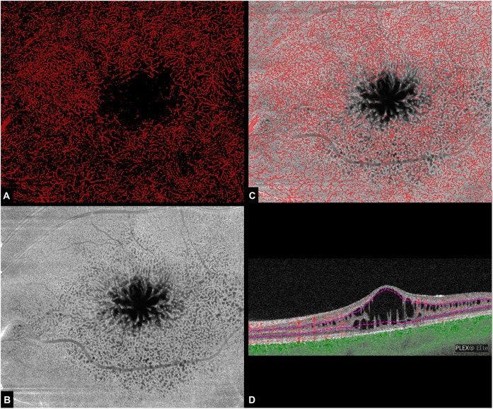
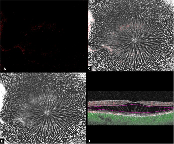
Methods: A case series of two patients, a 9-year-old male with XLR and a 58-year-old woman with SNIFR were imaged with swept-source optical coherence tomography angiography (SS-OCTA; PLEX Elite 900, Carl Zeiss Meditec, Inc, Dublin, CA). Automated segmentation was manually adjusted to include the areas of retinoschisis within en face flow and structural slabs. The flow data were binarized using ImageJ 1.51s (Wayne Rasband, National Institutes of Health, USA, http://imagej.nih.gov.ij ) and superimposed onto the structural slab.
Results: In the eye with XLR, OCTA flow data superimposed on the structural slab demonstrated flow signal within numerous bridging structures connecting the inner and outer plexiform layers containing the intermediate (ICP) and deep (DCP) capillary plexuses. In contrast, the same technique applied to the eye with SNIFR demonstrated an absence of flow signal in the cystic retinal spaces within Henle's fiber layer.
Conclusions: The vascular pattern of bridging vessels between the ICP and DCP is closely related to the structural "retinoschisis" pattern of XLR and appears to be structurally different from that seen in SNIFR. Moreover, the connecting vessels appear to be highly represented and regularly distributed, thereby supporting a serial arrangement of the retinal capillary plexuses within the perifoveal macula.
Conflict of interest statement
KBF is a consultant to Genentech, Allergan, Optos, Optovue, Zeiss, Heidelberg Engineering, and Novartis. He receives research funding from Genentech/Roche. JS is a consultant for Topcon, Diopsys, and INNOVA. The remaining authors declare that they have no conflict of interest.
Figures
Comment in
-
Vascular anatomy and its relationship to pathology in retinoschisis.Eye (Lond). 2019 May;33(5):693-694. doi: 10.1038/s41433-018-0298-6. Epub 2018 Dec 12. Eye (Lond). 2019. PMID: 30542065 Free PMC article. No abstract available.
References
-
- Orès Raphaëlle, Mohand-Said Saddek, Dhaenens Claire-Marie, Antonio Aline, Zeitz Christina, Augstburger Edouard, Andrieu Camille, Sahel José-Alain, Audo Isabelle. Phenotypic Characteristics of a French Cohort of Patients with X-Linked Retinoschisis. Ophthalmology. 2018;125(10):1587–1596. doi: 10.1016/j.ophtha.2018.03.057. - DOI - PubMed
Publication types
MeSH terms
LinkOut - more resources
Full Text Sources
Research Materials
Miscellaneous
