

Overexpression of RALY promotes migration and predicts poor prognosis in hepatocellular carcinoma
- PMID: 30519104
- PMCID: PMC6235007
- DOI: 10.2147/CMAR.S182996
Overexpression of RALY promotes migration and predicts poor prognosis in hepatocellular carcinoma
Abstract
Introduction: RALY plays a critical role in promoting invasiveness and is associated with poor prognosis in different types of cancers. However, the prognostic value of RALY and its precise role in hepatocellular carcinoma (HCC) remain unknown.
Materials and methods: We detected the expression of RALY in 127 clinical HCC tissue samples and seven HCC cell lines by immunohistochemical staining and Western blotting. The prognostic value of RALY expression was assessed using the Kaplan-Meier method. The expression and prognostic value of RALY were also studied by bioinformatics analysis of data from the Gene Expression Omnibus and The Cancer Genome Atlas. The biological influence of RALY on HCC cell lines was studied using proliferation, transwell migration, and invasion assays in vitro.
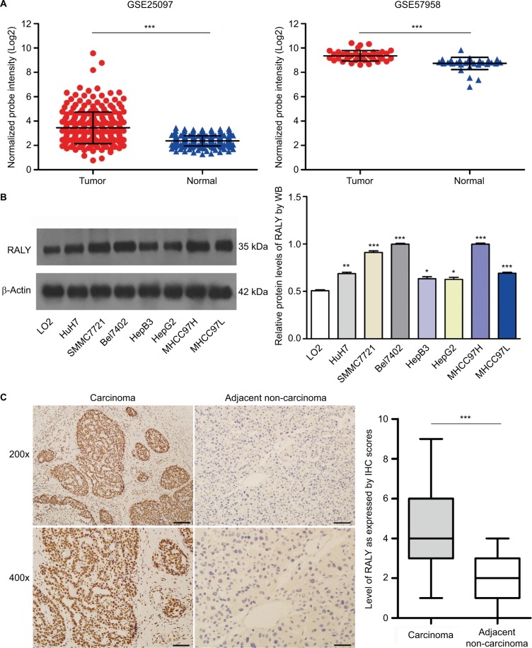
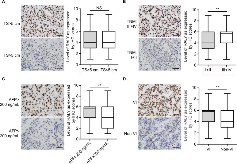
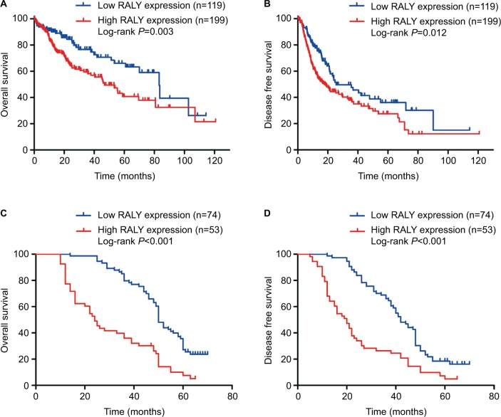
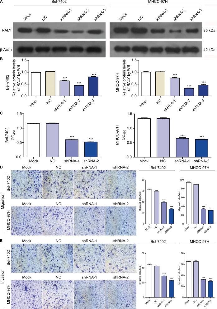
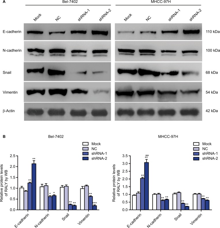
Results: The expression of RALY in HCC tissues was significantly higher than that in adjacent normal liver tissues. Abnormally high expression of RALY was associated with tumor size (P=0.031), TNM stage (P=0.026), presurgical serum AFP levels (P=0.025), and vascular invasion (P=0.001). Kaplan-Meier analysis demonstrated that higher expression of RALY correlated with poorer overall survival and disease-free survival in HCC patients. High RALY expression was an independent adverse prognostic factor for overall survival (HR =2.559, 95% CI: 1.710-3.827, P<0.001) and disease-free survival (HR =2.053, 95% CI: 1.384-3.047, P<0.001) in HCC. Moreover, knockdown of RALY expression using a specific shRNA suppressed the proliferation, migration, and invasion capabilities of HCC cells in vitro. Knockdown of RALY expression in HCC cell lines resulted in upregulation of E-cadherin and downregulation of N-cadherin, vimentin, and snail.
Conclusion: Taken together, our results indicate that RALY represents a biomarker for the prognosis of patients with HCC and highlight the importance of RALY as an oncogene in HCC.
Keywords: RALY; hepatocellular carcinoma; invasion; migration.
Conflict of interest statement
Disclosure The authors report no conflicts of interest in this work.
Figures
Similar articles
-
Neuron-glial antigen 2 overexpression in hepatocellular carcinoma predicts poor prognosis.World J Gastroenterol. 2015 Jun 7;21(21):6649-59. doi: 10.3748/wjg.v21.i21.6649. World J Gastroenterol. 2015. PMID: 26074703 Free PMC article.
-
Oncogene Delta/Notch-Like EGF-Related Receptor Promotes Cell Proliferation, Invasion, and Migration in Hepatocellular Carcinoma and Predicts a Poor Prognosis.Cancer Biother Radiopharm. 2018 Nov;33(9):380-386. doi: 10.1089/cbr.2018.2460. Epub 2018 Sep 20. Cancer Biother Radiopharm. 2018. PMID: 30235011 Free PMC article.
-
Long non-coding RNA SNHG20 predicts a poor prognosis for HCC and promotes cell invasion by regulating the epithelial-to-mesenchymal transition.Biomed Pharmacother. 2017 May;89:857-863. doi: 10.1016/j.biopha.2017.01.011. Epub 2017 Mar 6. Biomed Pharmacother. 2017. PMID: 28282787
-
FoxM1 overexpression promotes epithelial-mesenchymal transition and metastasis of hepatocellular carcinoma.World J Gastroenterol. 2015 Jan 7;21(1):196-213. doi: 10.3748/wjg.v21.i1.196. World J Gastroenterol. 2015. PMID: 25574092 Free PMC article.
-
Roles of diencephalon/mesencephalon homeobox 1 in the development and prognosis of hepatocellular carcinoma.Ann Hepatol. 2021 Sep-Oct;24:100314. doi: 10.1016/j.aohep.2021.100314. Epub 2021 Jan 29. Ann Hepatol. 2021. PMID: 33524552
Cited by
-
O-GlcNAcylated RALY Contributes to Hepatocellular Carcinoma Cells Proliferation by Regulating USP22 mRNA Nuclear Export.Int J Biol Sci. 2024 Jul 1;20(9):3675-3690. doi: 10.7150/ijbs.97397. eCollection 2024. Int J Biol Sci. 2024. PMID: 38993567 Free PMC article.
-
RALY regulate the proliferation and expression of immune/inflammatory response genes via alternative splicing of FOS.Genes Immun. 2022 Dec;23(8):246-254. doi: 10.1038/s41435-022-00178-4. Epub 2022 Aug 8. Genes Immun. 2022. PMID: 35941292 Free PMC article.
-
Transposon Insertion Mutagenesis in Mice for Modeling Human Cancers: Critical Insights Gained and New Opportunities.Int J Mol Sci. 2020 Feb 10;21(3):1172. doi: 10.3390/ijms21031172. Int J Mol Sci. 2020. PMID: 32050713 Free PMC article. Review.
-
Over-expression of RALYL suppresses the progression of ovarian clear cell carcinoma through inhibiting MAPK and CDH1 signaling pathways.Int J Med Sci. 2021 Jan 1;18(3):785-791. doi: 10.7150/ijms.51488. eCollection 2021. Int J Med Sci. 2021. PMID: 33437214 Free PMC article.
-
Proteomic analysis of hypopharyngeal and laryngeal squamous cell carcinoma sheds light on differences in survival.Sci Rep. 2020 Nov 10;10(1):19459. doi: 10.1038/s41598-020-76626-w. Sci Rep. 2020. PMID: 33173143 Free PMC article.
References
-
- Fan ST, Mau Lo C, Poon RT, et al. Continuous improvement of survival outcomes of resection of hepatocellular carcinoma: a 20-year experience. Ann Surg. 2011;253(4):745–758. - PubMed
-
- Lei Z, Li J, Wu D, et al. Nomogram for preoperative estimation of microvascular invasion risk in hepatitis B virus-related hepatocellular carcinoma within the Milan criteria. JAMA Surg. 2016;151(4):356–363. - PubMed
-
- Jiang L, Yan Q, Fang S, et al. Calcium-binding protein 39 promotes hepatocellular carcinoma growth and metastasis by activating extracellular signal-regulated kinase signaling pathway. Hepatology. 2017;66(5):1529–1545. - PubMed
LinkOut - more resources
Full Text Sources
Research Materials
Miscellaneous
