Review
. 2018 Nov 21:9:1630.
doi: 10.3389/fphys.2018.01630. eCollection 2018.

Current Perspectives on Tooth Implantation, Attachment, and Replacement in Amniota

Affiliations
Review

Current Perspectives on Tooth Implantation, Attachment, and Replacement in Amniota

Thomas J C Bertin et al. Front Physiol. .

Abstract

Teeth and dentitions contain many morphological characters which give them a particularly important weight in comparative anatomy, systematics, physiology and ecology. As teeth are organs that contain the hardest mineralized tissues vertebrates can produce, their fossil remains are abundant and the study of their anatomy in fossil specimens is of major importance in evolutionary biology. Comparative anatomy has long favored studies of dental characters rather than features associated with tooth attachment and implantation. Here we review a large part of the historical and modern work on the attachment, implantation and replacement of teeth in Amniota. We propose synthetic definitions or redefinitions of most commonly used terms, some of which have led to confusion and conflation of terminology. In particular, there has long been much conflation between dental implantation that strictly concerns the geometrical aspects of the tooth-bone interface, and the nature of the dental attachment, which mostly concerns the histological features occurring at this interface. A second aim of this work was to evaluate the diversity of tooth attachment, implantation and replacement in extant and extinct amniotes in order to derive hypothetical evolutionary trends in these different dental traits over time. Continuous dental replacement prevails within amniotes, replacement being drastically modified only in Mammalia and when dental implantation is acrodont. By comparison, dental implantation frequently and rapidly changes at various taxonomic scales and is often homoplastic. This contrasts with the conservatism in the identity of the tooth attachment tissues (cementum, periodontal ligament, and alveolar bone), which were already present in the earliest known amniotes. Because the study of dental attachment requires invasive histological investigations, this trait is least documented and therefore its evolutionary history is currently poorly understood. Finally, it is essential to go on collecting data from all groups of amniotes in order to better understand and consequently better define dental characters.

Keywords: acrodonty; amniota; evolution; periodontium; pleurodonty; thecodonty; tooth implantation; tooth replacement.


Figures

FIGURE 1
FIGURE 1
The amniote periodontium and tooth tissue organization. (A) Labio-lingual section of a tooth with a thecodont implantation and gomphosis attachment. (B) Labio-lingual section of a tooth with a pleurodont implantation and ankylosed attachment. Modified from (LeBlanc and Reisz, 2013).
FIGURE 2
FIGURE 2
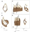
Tooth attachment illustrated by labio-lingual sections of mandibles in various species. (A) Gomphosis attachment associated with a thecodont implantation in Crocodylus niloticus. Note that the tooth and the bone are not in direct contact, leaving a periodontal space (ps). As the limit between alveolar bone and jaw bone is difficult to define, it is drawn as blurred. (B) Ankylosis attachment associated with a subthecodont implantation in Tupinambis teguxin (specimen MNHN 1967-96). The tooth is fused to a mineralized attachment tissue, leaving no periodontal space. Note the presence of a replacement pit (rp). (C) Tooth newly attached in Tupinambis teguxin (same specimen). The “attachment bone” is mineralized, which makes it more clearly distinguishable from the dentary bone. (D) Ankylosis attachment associated with a pleurodont implantation in Cyclura cornuta (specimen MNHN 1919-45). Scale bar is 2 mm.
FIGURE 3
FIGURE 3
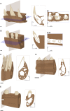
Implantation geometry illustrated by 3D portions of maxilla (A,B) and mandible (C–E) associated with virtual sections. (A) Thecodont implantation in the Nile crocodile (Crocodylus niloticus). (B) Aulacodont implantation in the Porpoise (Phocoena sp., specimen ENSL agSVSTUA 024181). (C) Pleurodont implantation in the Green iguana (Iguana iguana, specimen MNHN 1939-523). (D) Acrodont implantation in the Graceful chameleon (Chamaeleo gracilis, specimen MNHN 1942-114). (E) Subthecodont implantation in the Tegu (Tupinambis teguxin, specimen MNHN 1967-96). Cs, coronal side; Ls, lingual side; Ms, mesial side. Scale bar is 1 mm.
FIGURE 4
FIGURE 4
Tooth replacement illustrated by labio-lingual sections and perspective views of mandibles in various species. n, n+1, and n+2 indicate which generation teeth belong. (A) Diphyodonty in the rabbit (Oryctolagus cunniculus). (B) Polyphyodonty in the Nile crocodile (Crocodylus niloticus). (C) Labio-mesial replacement in Zamenis sp. (specimen MNHN 1969-789). (D) Labio-vertical replacement in Lacerta viridis (MNHN 1887-813). Coronal sections correspond to the gray section plan. Scale bar is 0.5 mm.
FIGURE 5
FIGURE 5
Implantation geometries placed next to the phylogenetic tree of squamates modified from (Pyron, 2016; Zheng and Wiens, 2016). Specimens illustrated from top to bottom are: Hemitheconyx caudicinctus (MNHN 1943-150); Eumeces algeriensis (MNHN 1886-343); Tupinambis teguxin (MNHN 1967-96); Amphisbaena alba (MNHN 1943-137); Lacerta viridis (MNHN 1887-813); Chamaeleo gracilis (MNHN 1942-114); Draco fimbriatus (MNHN 1887-880); Cyclura cornuta (MNHN 1919-45); Ophisaurus ventralis (MNHN 1943-143); Varanus niloticus (MNHN 1921-260); Cylindrophis rufus (MNHN 1869-779); Python sebae (MNHN 1953-155); Vipera aspis (MNHN 1869-855); Zamenis sp. (MNHN 1869-789).
FIGURE 6
FIGURE 6
Path of the replacement tooth in Squamata. (A) Labio-vertical path in Iguana iguana (specimen MNHN 1939-523). (B) Intermediate path between (A,C) in Ophiodes striatus (specimen MNHN 1943-142). (C) Labio-mesial path in Zamenis sp. (specimen MNHN 1969-789). Scale bar is 0.4 mm.
FIGURE 7
FIGURE 7
Dental features among early amniotes. Cladogram presenting some of the groups of stem or early Amniota. The dental features associated with each group are the assumed ancestral state for the groups or the described states for the genera (in bold). Other states that have been described in the groups are indicated with a smaller font. Implantation = geometry of implantation (S, subthecodonty; T, thecodonty; P, pleurodonty; A, acrodonty; Au, aulacodonty) Attachment = nature of the attachment (A, ankylosis; G, gomphosis) Replacement = number of replacement generation (P, polyphyodont; D, diphyodont; M, monophyodont). From (Romer and Price, 1940; Edmund, 1960, 1969; Ewer, 1965; Dong, 1972; Evans, 1984; Whiteside, 1986; Laurin and Reisz, 1995; Laurin, 1996b; Small, 1997; Rieppel, 2001; Cabreira and Cisneros, 2009; Dyke and Kaiser, 2011; Macdougall and Modesto, 2011; Pretto et al., 2012; LeBlanc and Reisz, 2013, 2015; Benton et al., 2015; Sassoon et al., 2015; de Miguel Chaves et al., 2018).
FIGURE 8
FIGURE 8
Current knowledge of the nature of the attachment tissues nature in different extinct and extant species represented from available literature. (A) Crocodylus niloticus (Archosauria, Crocodilidae) attachment is gomphosis. Scale bar is 2 mm. (B) Diadectes (Diadectomorpha) (LeBlanc and Reisz, 2013). Attachment is ankylosis. Scale bar is 1 cm (C) Iguana iguana (Squamata, Iguanidae) (Luan et al., 2009). Attachment is ankylosis. Scale bar is 1.5 mm (D) Dinilysia (Squamata, Dinilysiidae) (Budney et al., 2006). Attachment is hinged through soft ligament. Scale bar is 1 mm. (E) Chamaeleo calyptratus (Squamata, Chamaeleonidae) (Buchtová et al., 2013). The attachment is ankylosis, Scale bar is 0.8 mm. (F) Platecarpus (Squamata, Mosasauridea) (Caldwell, 2007). Attachment is ankylosis. Scale bar is 2 cm (G) Platypterygius, (Ichthyosauria) (Maxwell et al., 2011b). Attachment is gomphosis. The cellular cementum is vascularized (osteocementum). The panels (A’–G’) correspond to a magnification of the area framed on the panels (A–G), respectively. Scale bar is 1 cm.

References

    1. Andersson L., Blomlöf L., Lindskog S., Feiglin B., Hammarström L. (1984). Tooth ankylosis. Int. J. Oral Surg. 13 423–431. 10.1016/S0300-9785(84)80069-1 - DOI - PubMed
    1. Atrizadeh F., Kennedy J., Zander H. (1971). Ankylosis of teeth following thermal injury. J. Periodontal Res. 6 159–167. 10.1111/j.1600-0765.1971.tb00604.x - DOI - PubMed
    1. Auffenberg W. (1988). Gray’s Monitor Lizard. Gainsville FL: University Press of Florida.
    1. Bell G. L. (1997). “Chapter 11 - a phylogenetic revision of north american and adriatic mosasauroidea,” in Ancient Marine Reptiles, eds Callaway J. M., Nicholls E. L. (San Diego: Academic Press; ), 293–332.
    1. Bellairs A., Miles A. (1960). Apparent failure of tooth replacement in monitor lizards. Br. J. Herpet. 2 189–194.