Review
. 2018 Nov 21:9:1661.
doi: 10.3389/fphys.2018.01661. eCollection 2018.

Emerging Roles of the Membrane Potential: Action Beyond the Action Potential

Affiliations
Review

Emerging Roles of the Membrane Potential: Action Beyond the Action Potential

Lina Abdul Kadir et al. Front Physiol. .

Abstract

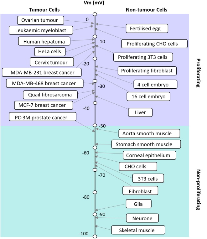
Whilst the phenomenon of an electrical resting membrane potential (RMP) is a central tenet of biology, it is nearly always discussed as a phenomenon that facilitates the propagation of action potentials in excitable tissue, muscle, and nerve. However, as ion channel research shifts beyond these tissues, it became clear that the RMP is a feature of virtually all cells studied. The RMP is maintained by the cell's compliment of ion channels. Transcriptome sequencing is increasingly revealing that equally rich compliments of ion channels exist in both excitable and non-excitable tissue. In this review, we discuss a range of critical roles that the RMP has in a variety of cell types beyond the action potential. Whereas most biologists would perceive that the RMP is primarily about excitability, the data show that in fact excitability is only a small part of it. Emerging evidence show that a dynamic membrane potential is critical for many other processes including cell cycle, cell-volume control, proliferation, muscle contraction (even in the absence of an action potential), and wound healing. Modulation of the RMP is therefore a potential target for many new drugs targeting a range of diseases and biological functions from cancer through to wound healing and is likely to be key to the development of successful stem cell therapies.

Keywords: blood pressure; cancer; ion channels; membrane potential; neurons; proliferation; resting membrane potential; stem cells.


Figures

FIGURE 1
FIGURE 1
Apparent correlation between the RMP and proliferative potential of cells. The RMP of tumor and non-tumor cells and their proliferation potential are shown. Modified with permission from the copyright holders Yang and Brackenbury (2013).
FIGURE 2
FIGURE 2
The electrochemical profile of the cochlea. (A) The structure of the human ear and a cross-section of the cochlea are illustrated in the upper panel. The tissue and cellular composition of the cochlea are illustrated in the lower panel; this shows the electrochemical properties of the endolymph and perilymph and the possible circulation of K+ ions between them. The boxed region in the lower panel is shown in (B). (B) Structural and cellular composition of the lateral wall of the cochlea depicting key ion channels and transporters that are thought to maintain the unidirectional K+ transport across the lateral wall and the endocochlear potential (EP) they produce are shown. (C) Membrane potential and [K+] of the lateral wall under physiological conditions. The top panel displays the membrane potential and [K+] averaged from multiple measurements. The membrane potential across the basolateral and apical surfaces of the syncytial layer, vSB and vSA, respectively, and the basolateral and apical surfaces of the marginal cell layer, vMB and vMA, respectively, are shown. The lower panel shows a representative trace of the measurement of the membrane potential and the [K+] in the cochlea of a live guinea pig. [Reproduced and modified with permission from the copyright holders the National Academy of Sciences (Nin et al., 2008) and Springer-Verlag Berlin Heidelberg (Yoshida et al., 2016)].

References

    1. Aguilar-Bryan L., Bryan J. (1999). Molecular biology of adenosine triphosphate-sensitive potassium channels. Endocr. Rev. 20 101–135. 10.1210/er.20.2.101 - DOI - PubMed
    1. Amigorena S., Choquet D., Teillaud J. L., Korn H., Fridman W. H. (1990). Ion channel blockers inhibit B cell activation at a precise stage of the G1 phase of the cell cycle. Possible involvement of K+ channels. J. Immunol. 144 2038–2045. - PubMed
    1. Ashcroft F. M., Harrison D. E., Ashcroft S. J. (1984). Glucose induces closure of single potassium channels in isolated rat pancreatic beta-cells. Nature 312 446–448. 10.1038/312446a0 - DOI - PubMed
    1. Ashcroft F. M., Rorsman P. (1990). Atp-sensitive K+ channels - a link between B-cell metabolism and insulin-secretion. Biochem. Soc. Trans. 18 109–111. 10.1042/bst0180109 - DOI - PubMed
    1. Ashmore J. (2008). Cochlear outer hair cell motility. Physiol. Rev. 88 173–210. 10.1152/physrev.00044.2006 - DOI - PubMed

LinkOut - more resources