. 2018 Nov 30:11:319.
doi: 10.1186/s13068-018-1321-7. eCollection 2018.

Cellulose-specific Type B carbohydrate binding modules: understanding oligomeric and non-crystalline substrate recognition mechanisms

Affiliations

Cellulose-specific Type B carbohydrate binding modules: understanding oligomeric and non-crystalline substrate recognition mechanisms

Abhishek A Kognole et al. Biotechnol Biofuels. .

Abstract

Background: Effective enzymatic degradation of crystalline polysaccharides requires a synergistic cocktail of hydrolytic enzymes tailored to the wide-ranging degree of substrate crystallinity. To accomplish this type of targeted carbohydrate recognition, nature produces multi-modular enzymes, having at least one catalytic domain appended to one or more carbohydrate binding modules (CBMs). The Type B CBM categorization encompasses several families (i.e., protein folds) of CBMs that are generally thought to selectively bind oligomeric polysaccharides; however, a subset of cellulose-specific CBM families (17 and 28) appear to bind non-crystalline cellulose more tightly than oligomers and in a manner that discriminates between surface topology.

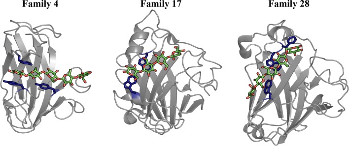
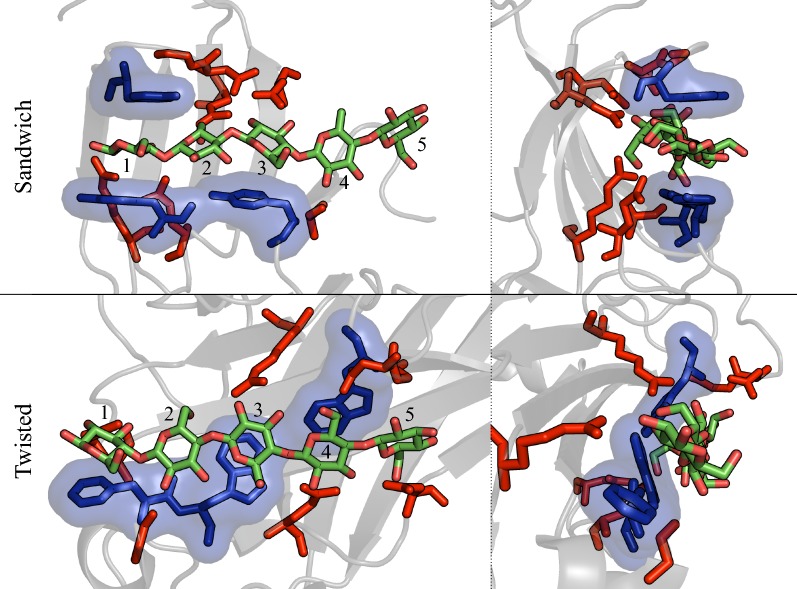
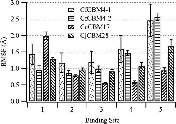
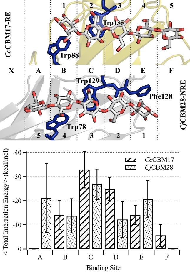
Results: To provide insight into this unexplained phenomenon, we investigated the molecular-level origins of oligomeric and non-crystalline carbohydrate recognition in cellulose-specific Type B CBMs using molecular dynamics (MD) simulation and free energy calculations. Examining two CBMs from three different families (4, 17, and 28), we describe how protein-ligand dynamics contribute to observed variations in binding affinity of oligomers within the same CBM family. Comparisons across the three CBM families identified factors leading to modified functionality prohibiting competitive binding, despite similarity in sequence and specificity. Using free energy perturbation with Hamiltonian replica exchange MD, we also examined the hypothesis that the open topology of the binding grooves in families 17 and 28 necessitates tight binding of an oligomer, while the more confined family 4 binding groove does not require the same degree of tight binding. Finally, we elucidated the mechanisms of non-crystalline carbohydrate recognition by modeling CBMs complexed with a partially decrystallized cellulose substrate. Molecular simulation provided structural and dynamic data for direct comparison to oligomeric modes of carbohydrate recognition, and umbrella sampling MD was used to determine ligand binding free energy. Comparing both protein-carbohydrate interactions and ligand binding free energies, which were in good agreement with experimental values, we confirmed the hypothesis that family 17 and 28 CBMs bind non-crystalline cellulose and oligomers with different affinities (i.e., high and low).

Conclusions: Our study provides an unprecedented level of insight into the complex solid and soluble carbohydrate substrate recognition mechanisms of Type B CBMs, the findings of which hold considerable promise for enhancing lignocellulosic biomass conversion technology and development of plant cell wall probes.

Keywords: Amorphous cellulose; Carbohydrate–aromatic stacking; Free energy perturbation; Multi-modular glycoside hydrolases; β-Sandwich fold.


Figures

Fig. 1
Fig. 1
CBMs (cartoon) from families 4, 17, and 28 with bound cello-oligomers (green and red sticks). Binding site aromatic residues are shown in a dark blue stick representation. The structures, Cellulomonas fimi CBM4-1, Clostridium cellulovorans CBM17, and Clostridium josui CBM28, were obtained from crystal structures with PDB IDs 1GU3, 1J84, and 3ACI, respectively. After structural alignment of the β-sandwich proteins, the family 4 and 17 CBM cello-oligomer is bound in the same direction, with the reducing end toward the left of the figure, whereas the family 28 CBM’s cello-oligomer is oriented in the opposite direction
Fig. 2
Fig. 2
Cartoon illustration of the protein–carbohydrate complexes modeled in this study. CBMs from family 17 and 28 were modeled with cellopentaose bound in a the crystallographic structure orientation and b with the reducing end of the pyranose ring at the opposite end of the groove from the structural orientation. c CBMs were also bi-directionally bound with partially decrystallized cellulose Iβ microfibrils, representative of non-crystalline cellulose substrates. RE reducing end, NRE non-reducing end
Fig. 3
Fig. 3
Differences in the two binding site architectures of family 4, 17, and 28 CBMs. Illustrated through hydrophobic interactions (dark blue sticks and transparent surface) and hydrogen bonding (red sticks) with the cellopentaose ligand (light green and red sticks). The front view (top left) and side view (top right) of the CfCBM4-1 binding site with bound cellopentaose clearly show the sandwich platform and deep cleft with one-sided hydrogen bonding of the ligand. The front view (bottom left) and side (bottom right) of the CjCBM28 binding site with bound cellopentaose show a twisted surface platform and shallow groove with hydrogen bonding partners available on both sides of ligand. The numbers on the cello-oligomeric ligand represents the binding subsite number
Fig. 4
Fig. 4
Root mean square fluctuation (RMSF) of cellopentaose. RMSF of the cellopentaose ligand from its average position in the clefts/grooves of representatives from family 4, 17, and 28 CBMs obtained from 250-ns MD simulation on a per-binding-subsite basis. Error was calculated from block averaging with block sizes of 2.5 ns
Fig. 5
Fig. 5
Average change in solvent accessible surface area (∆SASA) upon ligand binding. ΔSASA is calculated using VMD over the 250-ns MD simulation trajectories of each CBM–cellopentaose system to compare the difference between sandwich (lined pattern) and twisted (dotted pattern) platforms. The error bars represent the standard deviation of the mean
Fig. 6
Fig. 6
Alignment and comparison of the twisted platform binding sites. CcCBM17-RE and CjCBM28-NRE aligned with respect to the common pair of Trp residues (dark blue sticks) (top). The common naming of binding subsites used in this study (letters) is given between the alignment of the binding sites, and the original nomenclature (numbers) is given above and below the cartoon representations in the top panel. Average total interaction energy of the pyranose rings with the surrounding amino acid residues, on a per-subsite-basis, of CcCBM17-RE and CjCBM28-NRE calculated from the 250-ns trajectory (bottom). Error bars represent 1 standard deviation
Fig. 7
Fig. 7
Root mean square fluctuation (RMSF) of the ligand. RMSF of the cellopentaose ligand from its average position over 250-ns trajectories. The RMSF was calculated on a per-binding-subsite basis for all eight family 17 and 28 CBM–cellopentaose systems. The error bars represent the standard deviation of block averaged RMSFs with block sizes of 2.5 ns each
Fig. 8
Fig. 8
Total interaction energy between the substrate and each protein residue. Energies are averaged over the length of the MD simulations. The CcCBM17 (top) and BspCBM28 (bottom) residue numbers are shown along the x-axis. The simulation case label is given at left, four cases for each family 17 and 28 CBM. The magnitude of the interaction energy between a given residue and the bound ligand, as indicated in the case name, is shown in white-red-black gradient. Favorable interactions are more negative and, thus, darker/black. In cellopentaose binding, ligand direction does not affect CBM–cellopentaose interactions, as redundant protein residues along the binding groove maintain an association with cellopentaose. In non-crystalline cellulose binding, the CBM protein surface interacts with the surrounding carbohydrate, in both forward and reverse orientations, to enhance binding affinity; the new protein–carbohydrate interactions are unique for each CBM and each direction
Fig. 9
Fig. 9
Potential of mean force (PMF) in uncoupling a CcCBM17 and b BspCBM28 from non-crystalline cellulose. Umbrella sampling MD was conducted over 30 0.5-Å windows using the projection of the distance vector on the z-axis as the reaction coordinate

References

    1. Himmel ME, Ding SY, Johnson DK, Adney WS, Nimlos MR, Brady JW, Foust TD. Biomass recalcitrance: engineering plants and enzymes for biofuels production. Science. 2007;315:804–807. doi: 10.1126/science.1137016. - DOI - PubMed
    1. Sun Y, Cheng JY. Hydrolysis of lignocellulosic materials for ethanol production: a review. Bioresourc Technol. 2002;83:1–11. doi: 10.1016/S0960-8524(01)00212-7. - DOI - PubMed
    1. Boraston AB, Bolam DN, Gilbert HJ, Davies GJ. Carbohydrate-binding modules: fine-tuning polysaccharide recognition. Biochem J. 2004;382:769–781. doi: 10.1042/BJ20040892. - DOI - PMC - PubMed
    1. Shoseyov O, Shani Z, Levy I. Carbohydrate binding modules: biochemical properties and novel applications. Microbiol Mol Biol Rev. 2006;70:283-+. doi: 10.1128/MMBR.00028-05. - DOI - PMC - PubMed
    1. Linder M, Teeri TT. The roles and function of cellulose-binding domains. J Biotechnol. 1997;57:15–28. doi: 10.1016/S0168-1656(97)00087-4. - DOI - PubMed

LinkOut - more resources