

Promotion of Cell Proliferation through Inhibition of Cell Autophagy Signalling Pathway by Rab3IP is Restrained by MicroRNA-532-3p in Gastric Cancer
- PMID: 30519341
- PMCID: PMC6277663
- DOI: 10.7150/jca.27533
Promotion of Cell Proliferation through Inhibition of Cell Autophagy Signalling Pathway by Rab3IP is Restrained by MicroRNA-532-3p in Gastric Cancer
Abstract
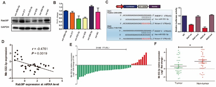
Background: RAB3A-interacting protein (Rab3IP) is known to be involved in cancer; however, its function during the proliferation of gastric cancer (GC) cells remains unknown. Therefore, this study aimed to explore the potential function of Rab3IP in GC. Methods: The expression of Rab3IP and its clinical pathology value were determined by quantitative real-time PCR and immunohistochemistry. Rab3IP (knockdown and overexpression) and light chain 3 (LC3) lentiviruses were transfected into GC cells, and cell proliferation was measured using cell counting kit-8, plate clone formation, flow cytometry, and tumorigenesis assays. Cell autophagy was measured using a confocal laser scanning microscope and by western blotting. Luciferase reporter assay was performed to analyse the regulation of Rab3IP by microRNA-532-3p (miR-532-3p). Results: Overexpression of Rab3IP in GC samples enhanced the cell proliferation ability, but decreased the number of autophagosomes and expression of LC3-II and sequestosome-1 (SQSTM1 or p62) markers. Furthermore, we found that miR-532-3p can bind to the 3'UTR region of RAB3IP and inhibit the proliferation ability of GC cells. Further, the expression of miR-532-3p negatively correlated with that of Rab3IP. Conclusions: Our study elucidates the central role of Rab3IP in inducing proliferation of GC cells through its involvement in autophagy. miR-532-3p directly targets Rab3IP and represses its function, thereby demonstrating a novel regulatory link in GC.
Keywords: Rab3IP; cell autophagy; cell proliferation; gastric cancer; miR-532-3p.
Conflict of interest statement
Competing Interests: The authors have declared that no competing interest exists.
Figures


Similar articles
-
18β-glycyrrhetinic acid promotes gastric cancer cell autophagy and inhibits proliferation by regulating miR-328-3p/signal transducer and activator of transcription 3.World J Gastroenterol. 2023 Jul 21;29(27):4317-4333. doi: 10.3748/wjg.v29.i27.4317. World J Gastroenterol. 2023. PMID: 37545635 Free PMC article.
-
Long noncoding RNA NEAT1 knockdown inhibits MPP+-induced apoptosis, inflammation and cytotoxicity in SK-N-SH cells by regulating miR-212-5p/RAB3IP axis.Neurosci Lett. 2020 Jul 13;731:135060. doi: 10.1016/j.neulet.2020.135060. Epub 2020 May 19. Neurosci Lett. 2020. PMID: 32442477
-
MicroRNA-101-3p regulates gastric cancer cell proliferation, invasion and apoptosis by targeting PIM 1 expression.Cell Mol Biol (Noisy-le-grand). 2019 Sep 30;65(7):118-122. Cell Mol Biol (Noisy-le-grand). 2019. PMID: 31880528
-
miR-27b-3p suppresses cell proliferation through targeting receptor tyrosine kinase like orphan receptor 1 in gastric cancer.J Exp Clin Cancer Res. 2015 Nov 14;34:139. doi: 10.1186/s13046-015-0253-3. J Exp Clin Cancer Res. 2015. PMID: 26576539 Free PMC article.
-
A novel long non-coding RNA XLOC_004787, is associated with migration and promotes cancer cell proliferation by downregulating mir-203a-3p in gastric cancer.BMC Gastroenterol. 2023 Aug 12;23(1):280. doi: 10.1186/s12876-023-02912-2. BMC Gastroenterol. 2023. PMID: 37573302 Free PMC article.
Cited by
-
Ectopic Expression of miR-532-3p Suppresses Bone Metastasis of Prostate Cancer Cells via Inactivating NF-κB Signaling.Mol Ther Oncolytics. 2020 Apr 7;17:267-277. doi: 10.1016/j.omto.2020.03.024. eCollection 2020 Jun 26. Mol Ther Oncolytics. 2020. PMID: 32368615 Free PMC article.
-
miR-140-3p is involved in the occurrence and metastasis of gastric cancer by regulating the stability of FAM83B.Cancer Cell Int. 2021 Oct 16;21(1):537. doi: 10.1186/s12935-021-02245-8. Cancer Cell Int. 2021. PMID: 34656115 Free PMC article.
-
Vitamin D modulation and microRNAs in gastric cancer: prognostic and therapeutic role.Transl Cancer Res. 2021 Jun;10(6):3111-3127. doi: 10.21037/tcr-20-2813. Transl Cancer Res. 2021. PMID: 35116620 Free PMC article. Review.
-
Facing Cell Autophagy in Gastric Cancer - What Do We Know so Far?Int J Gen Med. 2021 May 3;14:1647-1659. doi: 10.2147/IJGM.S298705. eCollection 2021. Int J Gen Med. 2021. PMID: 33976565 Free PMC article. Review.
-
Potential Therapeutic Action of Autophagy in Gastric Cancer Managements: Novel Treatment Strategies and Pharmacological Interventions.Front Pharmacol. 2022 Jan 28;12:813703. doi: 10.3389/fphar.2021.813703. eCollection 2021. Front Pharmacol. 2022. PMID: 35153766 Free PMC article. Review.
References
-
- Torre LA, Bray F, Siegel RL, Ferlay J, Lortet-Tieulent J, Jemal A. Global cancer statistics, 2012. CA Cancer J Clin. 2015;65:87–108. - PubMed
-
- Yamashita K, Sakuramoto S, Watanabe M. Genomic and epigenetic profiles of gastric cancer: Potential diagnostic and therapeutic applications. Surg Today. 2011;41:24–38. - PubMed
-
- Kelly EE, Horgan CP, Goud B, McCaffrey MW. The Rab family of proteins: 25 years on. Biochem Soc Trans. 2012;40:1337–1347. - PubMed
LinkOut - more resources
Full Text Sources
Other Literature Sources
Miscellaneous
