

Stroke: The past, present and future
- PMID: 30519643
- PMCID: PMC6276980
- DOI: 10.1177/2398212818810689
Stroke: The past, present and future
Abstract
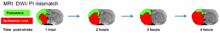
Since the inception of the British Neuroscience Association, there have been major advances in our knowledge of the mechanistic basis for stroke-induced brain damage. Identification of the ischaemic cascade led to the development of hundreds of new drugs, many showing efficacy in preclinical (animal-based) studies. None of these drugs has yet translated to a successful stroke treatment, current therapy being limited to thrombolysis/thrombectomy. However, this translational failure has led to significant improvements in the quality of animal-based stroke research, with the refinement of rodent models, introduction of new technologies (e.g. transgenics, in vivo brain imaging) and improvements in study design (e.g. STAIR, ARRIVE and IMPROVE guidelines). This has run in parallel with advances in clinical diagnostic imaging for detection of ischaemic versus haemorrhagic stroke, differentiating penumbra from ischaemic core, and improved clinical trial design. These preclinical and clinical advances represent the foundation for successful translation from the bench to the bedside in the near future.
Keywords: Stroke; brain injury; cerebral ischaemia; preclinical.
Conflict of interest statement
Declaration of conflicting interests The author(s) declared no potential conflicts of interest with respect to the research, authorship and/or publication of this article.
Figures
References
-
- Albanese V, Tommasino C, Spadaro A, et al. (1980) A transbasisphenoidal approach for selective occlusion of the middle cerebral artery in rats. Experientia 36(11): 1302–1304. - PubMed
-
- Astrup J, Siesjö BK, Symon L. (1981) Thresholds in cerebral ischemia – The ischemic penumbra. Stroke 12(6): 723–725. - PubMed
Grants and funding
LinkOut - more resources
Full Text Sources
Miscellaneous
