. 2018 Dec;23(12):1-4.
doi: 10.1117/1.JBO.23.12.121617.

Transvaginal fast-scanning optical-resolution photoacoustic endoscopy

Affiliations

Transvaginal fast-scanning optical-resolution photoacoustic endoscopy

Yuan Qu et al. J Biomed Opt. 2018 Dec.

Abstract

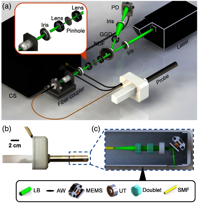
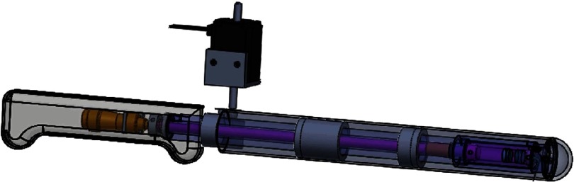
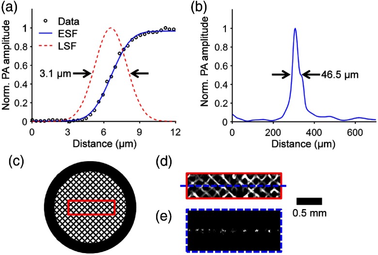
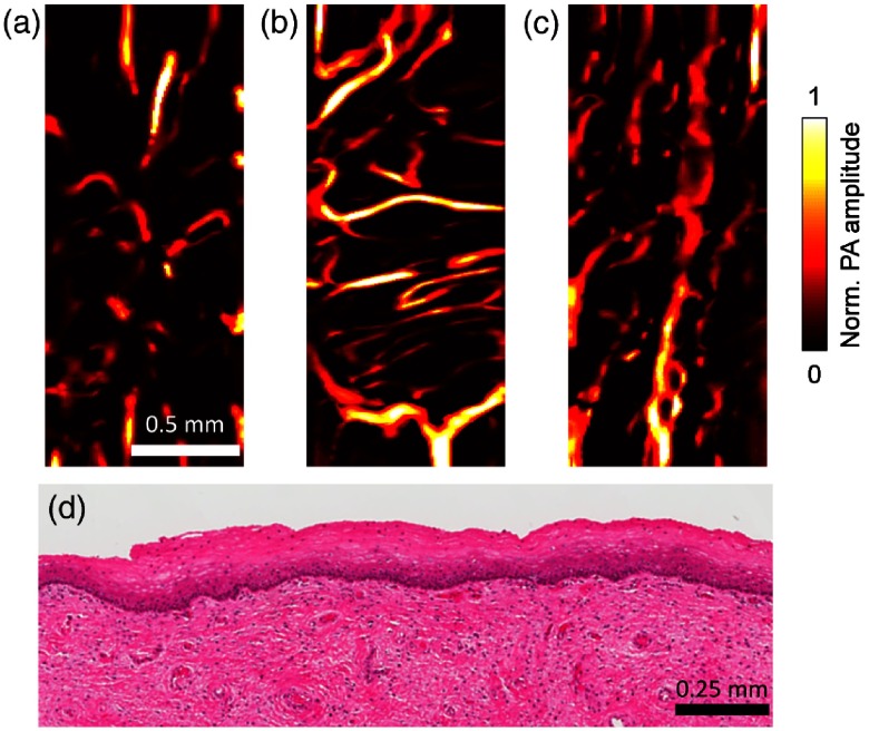
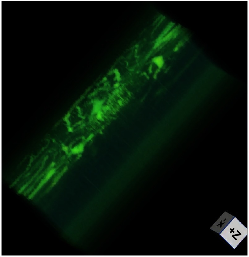
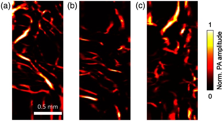
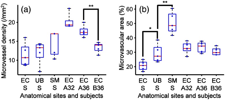
Photoacoustic endoscopy offers in vivo examination of the visceral tissue using endogenous contrast, but its typical B-scan rate is ∼10 Hz, restricted by the speed of the scanning unit and the laser pulse repetition rate. Here, we present a transvaginal fast-scanning optical-resolution photoacoustic endoscope with a 250-Hz B-scan rate over a 3-mm scanning range. Using this modality, we not only illustrated the morphological differences of vasculatures among the human ectocervix, uterine body, and sublingual mucosa but also showed the longitudinal and cross-sectional differences of cervical vasculatures in pregnant women. This technology is promising for screening the visceral pathological changes associated with angiogenesis.

Keywords: cervical imaging; fast scanning; microelectromechanical system scanning mirror; optical-resolution photoacoustic endoscopy.


Figures

Fig. 1
Fig. 1
Schematic of the fsOR-PAE probe and its peripheral systems. (a) Setup of fsOR-PAE. CS, control system; GGD, ground glass diffuser; NDF, variable neutral density filter; PD, photodetector. (b) Photograph of the fsOR-PAE probe. A linear actuator in the white housing drives the azimuth scanning. (c) Schematic of the acoustic-optical coaxial confocal alignment in the probe. MEMS drives the scanning parallel to the cylindrical axis. AW, acoustic wave; LB, laser beam; MEMS, microelectromechanical system scanning mirror; SMF, single-mode fiber; UT, ultrasonic transducer.
Fig. 2
Fig. 2
Scanning mechanism of the fsOR-PAE probe (Video 1, MP4, 3.78 MB [URL: https://doi.org/10.1117/1.JBO.23.12.121617.1]).
Fig. 3
Fig. 3
Characterization of the fsOR-PAE probe. (a) Lateral resolution test on a sharp edge. ESF, edge spread function; LSF, line spread function derived from ESF. (b) Axial resolution test on a tungsten wire. (c) Photograph of a metal grid. (d) Maximum amplitude projection image computed from the region enclosed by the red rectangle in (c). (e) B-scan image in the plane highlighted by the blue dashed line in (d).
Fig. 4
Fig. 4
Ex vivo fsOR-PAE images of (a) the human ectocervix, (b) the serosal layer of the uterine body, and (c) the sublingual mucosa. (d) Standard hematoxylin and eosin histology of the ectocervix conducted after fsOR-PAE imaging, showing no tissue damage.
Fig. 5
Fig. 5
Volume-rendered image (Video 2, MP4, 890 KB [URL: https://doi.org/10.1117/1.JBO.23.12.121617.2]).
Fig. 6
Fig. 6
In vivo fsOR-PAE images acquired from the first pregnant woman at (a) 32 and (b) 36 weeks of gestation. (c) In vivo fsOR-PAE image acquired from the second pregnant woman at 36 weeks of gestation.
Fig. 7
Fig. 7
Box plots for the histomorphological quantities calculated from the fsOR-PAE images. Five images were analyzed for each subject. (a) Microvessel density and (b) total microvascular area. EC A32, the ectocervix of the first patient at 32 weeks of gestation; EC A36, the ectocervix of the first patient at 36 weeks of gestation; EC B36, the ectocervix of the second patient at 36 weeks of gestation; EC S, the ectocervix specimen; SM S, the sublingual mucosa specimen; UB S, the uterine body specimen. *P<0.05, **P<0.01.

References

    1. Leslie M., “Tumors’ do-it-yourself blood vessels,” Science 352, 1381–1383 (2016).SCIEAS10.1126/science.352.6292.1381 - DOI - PubMed
    1. Li L., et al. , “Single-impulse panoramic photoacoustic computed tomography of small-animal whole-body dynamics at high spatiotemporal resolution,” Nat. Biomed. Eng. 1, 0071 (2017).10.1038/s41551-017-0071 - DOI - PMC - PubMed
    1. He Y., et al. , “In vivo label-free photoacoustic flow cytography and on-the-spot laser killing of single circulating melanoma cells,” Sci. Rep. 6, 39616 (2016).SRCEC310.1038/srep39616 - DOI - PMC - PubMed
    1. Carmeliet P., Jain R. K., “Molecular mechanisms and clinical applications of angiogenesis,” Nature 473, 298–307 (2011).10.1038/nature10144 - DOI - PMC - PubMed
    1. Sharma S., Sharma M. C., Sarkar C., “Morphology of angiogenesis in human cancer: a conceptual overview, histoprognostic perspective and significance of neoangiogenesis,” Histopathology 46, 481–489 (2005).HISTDD10.1111/his.2005.46.issue-5 - DOI - PubMed

Publication types