

Increased Vulnerability to Atrial Fibrillation Is Associated With Increased Susceptibility to Alternans in Old Sheep
- PMID: 30520673
- PMCID: PMC6405564
- DOI: 10.1161/JAHA.118.009972
Increased Vulnerability to Atrial Fibrillation Is Associated With Increased Susceptibility to Alternans in Old Sheep
Erratum in
-
Increased Vulnerability to Atrial Fibrillation Is Associated With Increased Susceptibility to Alternans in Old Sheep.J Am Heart Assoc. 2019 Dec 17;8(24):e04679. doi: 10.1161/JAHA.118.002331. Epub 2019 Dec 4. J Am Heart Assoc. 2019. PMID: 31795825 Free PMC article. No abstract available.
Abstract
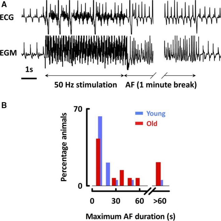
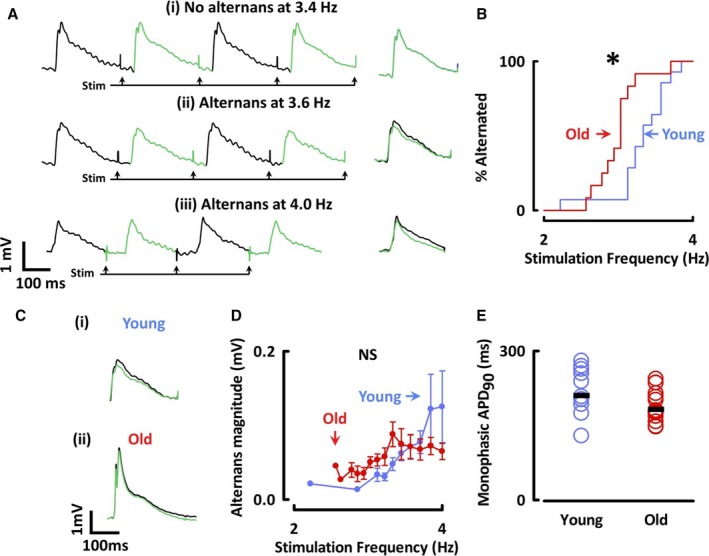
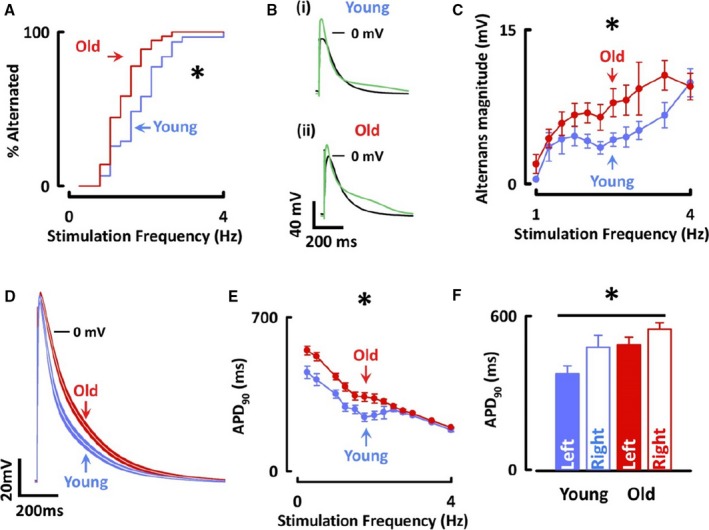
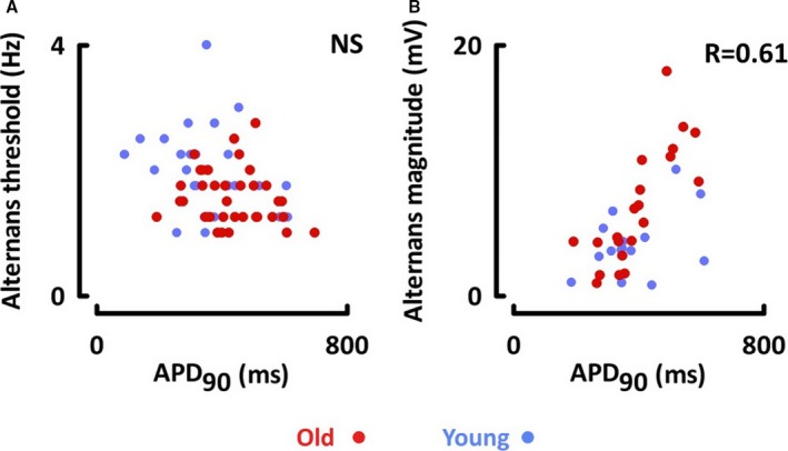
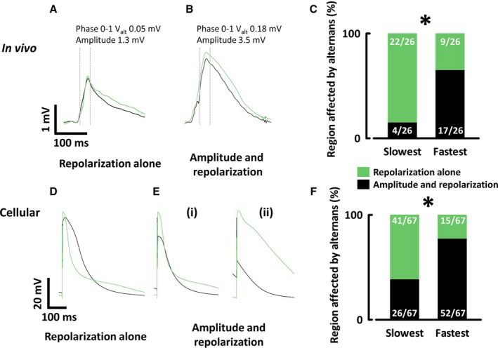
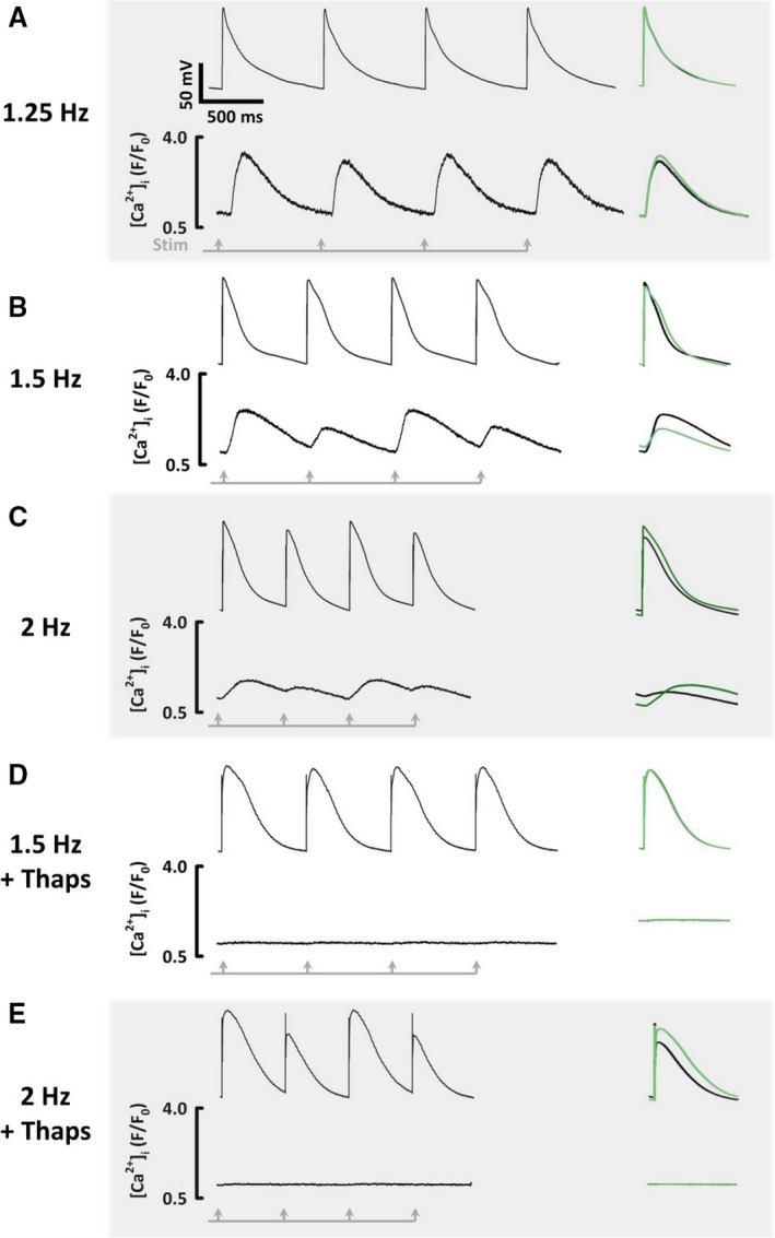
Background Atrial fibrillation ( AF ) is common in the elderly, but rare in the young; however, the changes that occur with age that promote AF are not fully understood. Action potential ( AP ) alternans may be involved in the initiation of AF . Using a translationally relevant model, we investigated whether age-associated atrial vulnerability to AF was associated with susceptibility to AP alternans. Methods and Results AF was induced in conscious young and old sheep using 50 Hz burst pacing. Old sheep were more vulnerable to AF . Monophasic and cellular AP s were recorded from the right atrium in vivo and from myocytes isolated from the left and right atrial appendages. AP alternans occurred at lower stimulation frequencies in old sheep than young in vivo (old, 3.0±0.1 Hz; young, 3.3±0.1 Hz; P<0.05) and in isolated myocytes (old, 1.6±0.1 Hz; young, 2.0±0.1 Hz; P<0.05). Simultaneous recordings of [Ca2+]i and membrane potential in myocytes showed that alternans of AP s and [Ca2+]i often occurred together. However, at low stimulation rates [Ca2+]i alternans could occur without AP alternans, whereas at high stimulation rates AP alternans could still be observed despite disabling Ca2+ cycling using thapsigargin. Conclusions We have shown, for the first time in a large mammalian model, that aging is associated with increased duration of AF and susceptibility to AP alternans. We suggest that instabilities in Ca2+ handling initiate alternans at low stimulation rates, but that AP restitution alone can sustain alternans at higher rates.
Keywords: action potential; aging; alternans; atrial fibrillation; calcium regulation.
Figures

References
-
- Go AS, Hylek EM, Phillips KA, Chang Y, Henault LE, Selby JV, Singer DE. Prevalence of diagnosed atrial fibrillation in adults: national implications for rhythm management and stroke prevention: the AnTicoagulation and Risk Factors in Atrial Fibrillation (ATRIA) Study. JAMA. 2001;285:2370–2375. - PubMed
-
- Schotten U, Verheule S, Kirchhof P, Goette A. Pathophysiological mechanisms of atrial fibrillation: a translational appraisal. Physiol Rev. 2011;91:265–325. - PubMed
-
- Verrier RL, Klingenheben T, Malik M, El‐Sherif N, Exner DV, Hohnloser SH, Ikeda T, Martinez JP, Narayan SM, Nieminen T, Rosenbaum DS. Microvolt T‐wave alternans physiological basis, methods of measurement, and clinical utility—consensus guideline by International Society for Holter and Noninvasive Electrocardiology. J Am Coll Cardiol. 2011;58:1309–1324. - PMC - PubMed
Publication types
MeSH terms
Substances
Grants and funding
- FS/12/57/29717/BHF_/British Heart Foundation/United Kingdom
- FS/12/34/29565/BHF_/British Heart Foundation/United Kingdom
- FS/09/002/26487/BHF_/British Heart Foundation/United Kingdom
- PG/12/89/29970/BHF_/British Heart Foundation/United Kingdom
- FS/17/54/33126/BHF_/British Heart Foundation/United Kingdom
LinkOut - more resources
Full Text Sources
Medical
Miscellaneous
