. 2018 Dec 6;13(12):e0208321.
doi: 10.1371/journal.pone.0208321. eCollection 2018.

Interleukin-6 inhibition of hERG underlies risk for acquired long QT in cardiac and systemic inflammation

Affiliations

Interleukin-6 inhibition of hERG underlies risk for acquired long QT in cardiac and systemic inflammation

Ademuyiwa S Aromolaran et al. PLoS One. .

Abstract

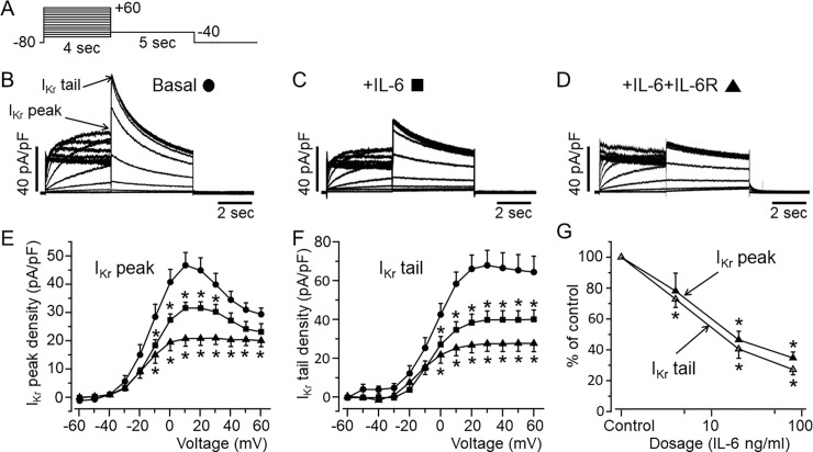
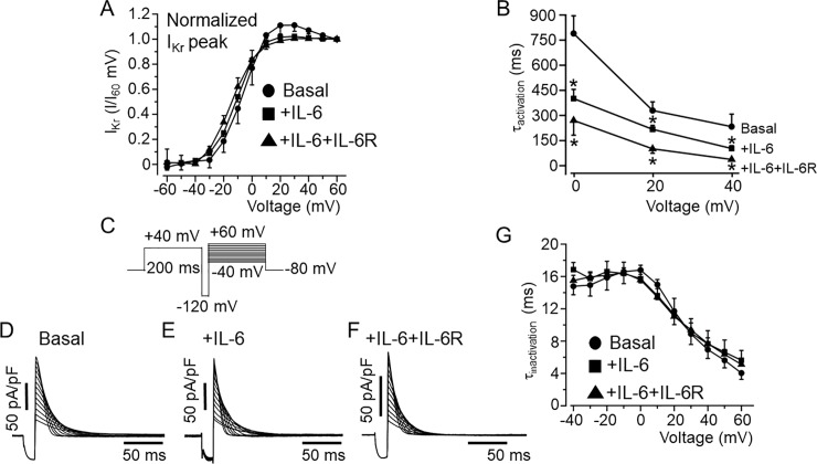
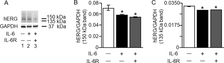
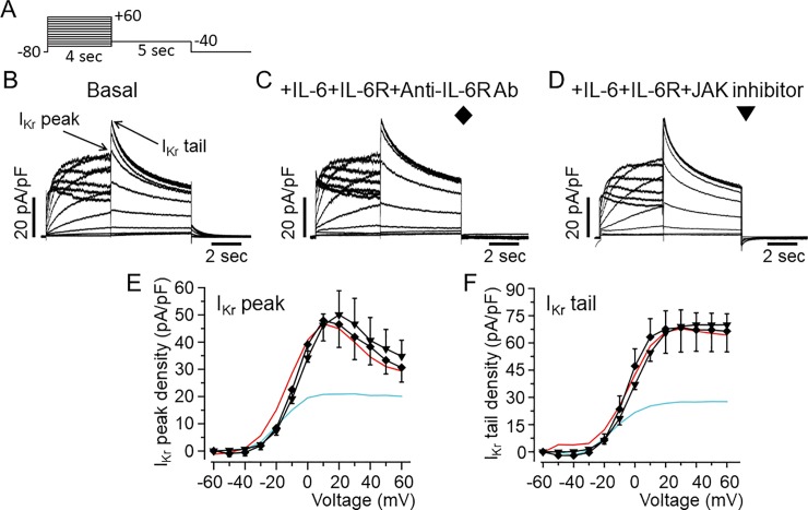
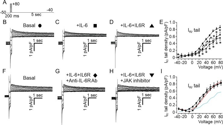
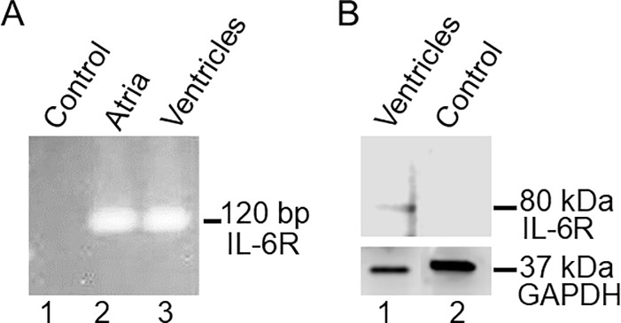
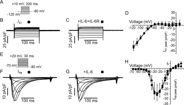
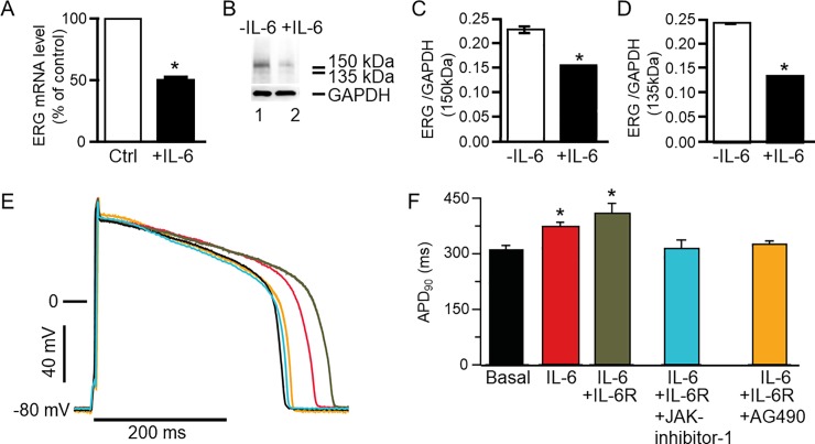
Increased proinflammatory interleukin-6 (IL-6) levels are associated with acquired long QT-syndrome (LQTS) in patients with systemic inflammation, leading to higher risks for life-threatening polymorphic ventricular tachycardia such as Torsades de Pointes. However, the functional and molecular mechanisms of this association are not known. In most cases of acquired LQTS, the target ion channel is the human ether-á-go-go-related gene (hERG) encoding the rapid component of the delayed rectifier K current, IKr, which plays a critical role in cardiac repolarization. Here, we tested the hypothesis that IL-6 may cause QT prolongation by suppressing IKr. Electrophysiological and biochemical assays were used to assess the impact of IL-6 on the functional expression of IKr in HEK293 cells and adult guinea-pig ventricular myocytes (AGPVM). In HEK293 cells, IL-6 alone or in combination with the soluble IL-6 receptor (IL-6R), produced a significant depression of IKr peak and tail current densities. Block of IL-6R or Janus kinase (JAK) reversed the inhibitory effects of IL-6 on IKr. In AGPVM, IL-6 prolonged action potential duration (APD) which was further prolonged in the presence of IL-6R. Similar to heterologous cells, IL-6 reduced endogenous guinea pig ERG channel mRNA and protein expression. The data are first to demonstrate that IL-6 inhibition of IKr and the resulting prolongation of APD is mediated via IL-6R and JAK pathway activation and forms the basis for the observed clinical QT interval prolongation. These novel findings may guide the development of targeted anti-arrhythmic therapeutic interventions in patients with LQTS and inflammatory disorders.


Conflict of interest statement

The authors have declared that no competing interests exist.

Figures

Fig 1
Fig 1. Functional effect of IL-6 on IKr in HEK-hERG cells.
A, Voltage-clamp protocol used to elicit IKr in HEK-hERG cells stable expressing hERG channel. B, Control hERG current. Arrows indicate hERG peak and tail currents. C, IKr currents measured in cells pre-treated with IL-6 alone or D, with IL-6+IL-6R. E, IKr peak and F, tail density-voltage- curves for control (circle, n = 21), IL-6 alone (square, n = 18) and IL-6+IL-6R (triangle, n = 9). G, The dose-response effects of IL-6+IL-6R on IKr peak and tail densities. Data were normalized to control and expressed as percentage of control.
Fig 2
Fig 2. Effects of IL-6 on IKr activation and inactivation kinetics in HEK-hERG cells.
A, Plots of IKr tail I/I+60mV for control and in the presence of IL-6 alone and IL-6+IL-6R. Compared to control IKr currents (circle, n = 21), the normalized tail currents show that IKr activates at more negative potentials in the presence IL-6 (square, n = 18) and IL-6+IL-6R (triangle, n = 9). B, Plots of activation time course (τactivation) and voltage relationship for basal IKr (circle, n = 21), and in the presence of IL-6 (square, n = 18) or IL-6+IL-6R (triangle, n = 9). The time course of inactivation (τinactivation) was examined using the protocol shown in C. D, representative IKr current traces recorded under basal conditions, and in the presence of IL-6 (E) or IL-6+IL-6R (F). G, graph of the voltage-dependence of the τinactivation during basal IKr (circle, n = 8), +IL-6 (square, n = 7), +IL-6+IL-6R (triangle, n = 5) conditions.
Fig 3
Fig 3. Effect of IL-6 on IKr protein expression in HEK-hERG cells.
Western blot analysis of the IKr channel hERG expression in HEK-hERG cells from control cells and in cells pre-treated for 40 min with IL-6 (20 ng/ml) alone or IL-6 (20 ng/ml)+IL-6R (25 ng/ml). A, compared to control hERG protein expression (Lane 1), exposure to IL-6 (20 ng/ml) reduced the expression of the 150 kDa and 135 kDa (Lane 2) hERG proteins. A further decrease in the bands was observed (Lane 3) with pretreatment with IL-6 (20 ng/ml) + IL-6R (25 ng/ml). Compared to untreated HEK-hERG cells (Open bar, B and C), normalized values of 150 kDa (B) and 135 kDa (C) hERG bands/GAPDH revealed significant down-regulation of the protein expression of hERG in the presence of IL-6 alone (Black Bars, B and C) or IL-6+IL-6R (Grey Bars, B and C). Equal amounts of protein were loaded in each lane. The 37 kDa bands represent GAPDH.
Fig 4
Fig 4. Inhibition of IKr by IL-6 is prevented by IL-6R and Janus Kinase blockade in HEK293 cells.
A, Voltage-clamp protocol used to generate IKr in HEK-hERG cells stably expressing hERG channel. B, Basal hERG currents. Arrows indicate hERG peak and tail currents. C, Representative IKr traces from HEK-hERG cells pretreated with IL-6+IL-6R in the presence of anti-IL-6R antibody (Ab). D, IKr currents measured in HEK-hERG cells pre-treated with IL-6+IL-6R and the JAK inhibitor-1. Plots of IKr peak current density-voltage (E) and tail current density-voltage curves (F) in the presence of anti-IL-6R antibody (n = 4) and the JAK inhibitor-1 (n = 4). IKr peak current and tail density-voltage curves for IKr measured in basal condition (Red line) and in cells pre-treated with IL-6+IL-6R (Cyan line) are shown for comparison.
Fig 5
Fig 5. Effects of IL-6 and IL-6+IL-6R on IKr in adult guinea-pig ventricular myocytes.
A, Voltage protocol used for evoking IKr in freshly isolated ventricular myocytes from adult guinea-pig heart. Tail current traces measured in the presence of 100 μM chromanol 293B and 5 μM nifedipine in control untreated cardiomyocyte (B, circle, n = 15), and myocytes pre-treated with IL-6 alone (C, square, n = 8) or IL-6 +IL-6R (D, upward triangle, n = 5). E, Population IKr tail density-voltage curves in basal, IL-6- and IL-6R-treated adult guinea-pig ventricular cardiomyocytes. F, Representative IKr traces in basal condition, (G) pre-treatment with IL-6+IL-6R in the presence of anti-IL-6R antibody (Ab) at 100 μg/ml or, H) a JAK inhibitor-I (5 μM). I, the mean IKr tail density-voltage curves show that the inhibitory effect of IL-6+IL-6R on IKr is reversed in the presence of anti-IL-6R antibody (diamond, n = 12) or JAK inhibitor I (downward triangle, n = 5). For visual comparison, data for control IKr (Red trace) and in the presence of IL-6+IL-6R (Cyan trace) in I.
Fig 6
Fig 6. Transcript and protein expression of IL-6R in guinea pig heart.
A, qRT-PCR assay for detection of IL-6R mRNA expression in guinea-pig heart. Compared to control (Lane 1), IL-6R (120 bp) mRNA expression was detected in both atria (Lane 2) and ventricles (Lane 3) consistent with an endogenous IL-6R expression in guinea pig heart. B, Western blot of IL-6R from guinea-pig ventricular cell lysates. The blot was probed with anti-IL6Rα H-7. The band around 80 kDa (Fig 6B, Lane 1) represents IL-6Rα which was absent when IL-6Rα Ab was pre-incubated with its own blocking peptide (Fig 6B, Lane 2).
Fig 7
Fig 7. IL-6 has no effect on IK1 or INa in guinea-pig ventricular myocytes.
A, Voltage protocol for activation of IK1. Representative IK1 traces for a control myocyte (B) and from an IL-6+IL-6R pre-treated myocyte (C). D, Population IK1 peak density-voltage curves show that IK1 was not altered in IL-6-IL-6R pre-treated myocytes (square, n = 4) compared to untreated myocytes (circle, n = 3) at all voltages between -120 mV to +10 mV. E, The voltage protocol for evoking INa. Representative traces showing that INa measured in control cardiomyocytes (F) are identical to INa recorded in myocytes pre-treated with IL-6 alone (G). H, pooled data for INa peak-voltage in untreated (circle, n = 6), and IL-6-treated myocytes (square, n = 6).
Fig 8
Fig 8. Effect of IL-6 on guinea pig ERG expression and action potential in ventricular myocytes.
A, qRT-PCR was used to determine guinea pig ether-á-go-go-related gene (ERG) mRNA expression in control ventricular myocytes and in myocytes pre-treated with IL-6 (20 ng/ml). B, Western blot assay of guinea pig ERG protein expression in control (Lane 1) and IL-6 pretreated (for 40 mins) myocytes (Lane 2). Values were normalized to the GAPDH signal and expressed as % of control or in the absence of IL-6. Each sample was analyzed in triplicate. 150 kDa and 135 kDa ERG bands were identified in lane 1 but were significantly less dense in lane 2 loaded with IL-6 (20 ng/ml) pretreated ventricular myocytes’ lysate. C, Comparison of the relative abundance of the 150 kDa ERG band in untreated and IL-6 treated myocytes. D, Comparison of the relative abundance of the 135 kDa ERG band in untreated and IL-treated myocytes. The 37 kDa bands represent GAPDH. E, Action potential waveforms recorded in control myocytes (basal conditions without IL-6, Black trace) and in the presence of IL-6 alone (Red trace), IL-6+IL-6R (Grey trace), IL-6+IL-6R+JAK inhibitor-1 (Cyan trace) and another JAK inhibitor AG490 (Orange trace). F, Pretreatment of myocytes with IL-6 and IL-6+IL-6R significantly prolonged APD90 and JAK inhibitor I and AG490 completely reversed the prolongation of the action potential.

References

    1. Yang S, Zheng R, Hu S, Ma Y, Choudhry MA, Messina JL, et al. Mechanism of cardiac depression after trauma-hemorrhage: increased cardiomyocyte IL-6 and effect of sex steroids on IL-6 regulation and cardiac function. American journal of physiology Heart and circulatory physiology. 2004;287(5):H2183–91. Epub 2004/10/12. 10.1152/ajpheart.00624.2003 . - DOI - PubMed
    1. Fontes JA, Rose NR, Cihakova D. The varying faces of IL-6: From cardiac protection to cardiac failure. Cytokine. 2015;74(1):62–8. Epub 2015/02/05. 10.1016/j.cyto.2014.12.024 ; PubMed Central PMCID: PMCPmc4677779. - DOI - PMC - PubMed
    1. Naka T, Nishimoto N, Kishimoto T. The paradigm of IL-6: from basic science to medicine. Arthritis Res. 2002;4 Suppl 3:S233–42. Epub 2002/07/12. 10.1186/ar565 ; PubMed Central PMCID: PMCPMC3240141. - DOI - PMC - PubMed
    1. Kurdi M, Booz GW. Can the protective actions of JAK-STAT in the heart be exploited therapeutically? Parsing the regulation of interleukin-6-type cytokine signaling. J Cardiovasc Pharmacol. 2007;50(2):126–41. Epub 2007/08/19. 10.1097/FJC.0b013e318068dd49 . - DOI - PubMed
    1. Lazzerini PE, Laghi-Pasini F, Bertolozzi I, Morozzi G, Lorenzini S, Simpatico A, et al. Systemic inflammation as a novel QT-prolonging risk factor in patients with torsades de pointes. Heart (British Cardiac Society). 2017. Epub 2017/05/12. 10.1136/heartjnl-2016-311079 . - DOI - PubMed

Publication types

MeSH terms